Start your free 3-day trial — no credit card required, full access included

Anatomy - Adnexectomy, laparoscopic, robot-assisted laparoscopy (DaVinci)

  1. Ovary (Ovarium)

    Ovary (Ovarium) 1
    Ovary (Ovarium) 2

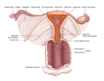
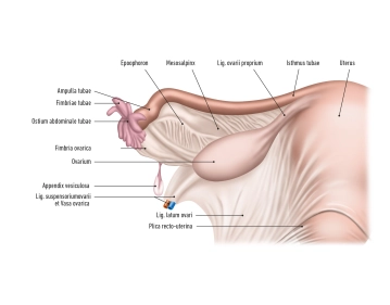
    The ovary (Ovarium) is located laterally in the pelvic region and runs in a vertical orientation. It has an inner surface facing the interior of the pelvis (Facies medialis/intestinalis) and an outer surface directed towards the lateral pelvic wall (Facies lateralis). The rounded free edge is referred to as the Margo liber, and at the anterior edge, the Margo mesovaricus, the mesovarium is attached, which is a structure of the broad ligament of the uterus (Lig. latum uteri).

    There are two poles:

    • Extremitas uterina
    • Extremitas tubaria

    The hilum of the ovary, where vessels and nerves enter, is located at the attachment of the connective tissue of the ovary, known as the mesovarium, at the Margo mesovaricus.

    The ovary is connected to the pelvic wall at the Extremitas tubaria by the suspensory ligament of the ovary. The ovarian vessels (Vasa ovarica), lymphatic vessels, and nerves run along this suspensory ligament.

    The proper ovarian ligament lies between the ovary (Extremitas uterina). This is located directly behind the tubal angle. The artery R. ovaricus of the uterine artery runs here.

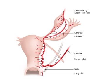
    The ovary is supplied by:

    • A. ovarica
    • Ramus ovaricus from the uterine artery.

    These arteries form anastomoses at the Margo mesovaricus and supply the ovary with blood.

    Veins transport the venous blood via the right ovarian vein to the inferior vena cava and via the left ovarian vein to the left renal vein. Part of the blood flows through the uterine plexus to the internal iliac vein.

    The ovary is located in the abdominal cavity (intraperitoneal) in the ovarian fossa. Here, in the retroperitoneum, run the obturator nerve, obturator vessels, and external iliac vessels. Medially, the umbilical artery and the uterine artery also run.

  2. The fallopian tube (Tuba uterina, Salpinx)

    The fallopian tube (Tuba uterina, Salpinx)

    The fallopian tube, also known as the uterine tube, is approximately 10-15 centimeters long and 2-5 millimeters thick. It runs intraperitoneally along the upper free edge of the broad ligament of the uterus (ligamentum latum uteri), from the corner of the uterus to the ovary. In the mesosalpinx, the blood vessels and nerves to the fallopian tube run, and additionally, the fallopian tube is attached to the ligamentum latum.

    The fallopian tube itself can be divided into different sections:

    • Infundibulum of the uterine tube (+ abdominal ostium of the uterine tube)
    • Fimbriae of the uterine tube,
    • Ampulla of the uterine tube
    • Isthmus of the uterine tube
    • Uterine part.

    The blood supply of the uterine tube is provided by small branches (Rr. tubarii) from the arteries A. ovarica and A. uterina, which run in the mesosalpinx and anastomose with each other. Veins accompany the arteries and drain into the V. ovarica and into the uterine/uterovaginal plexus.

  3. The uterus

    The uterus

    The uterus, also known as the womb, is a hollow, muscular organ in the female body that plays an important role during pregnancy and childbirth.

    The uterus consists of:

    • the body (Corpus uteri),
    • the isthmus uteri
    • the cervix (Cervix uteri)

    The average length of the uterus is about 7.5 cm, the width 4 cm, and the thickness 2.5 cm.

    The fundus uteri represents the upper section of the uterus, located above the entry points of the fallopian tubes. Its lateral walls are connected with the broad ligament (Ligamentum latum uteri). At the lateral edges of the uterus, where the fallopian tubes enter the uterus, the tubal angle is formed. From the tubal angle, the proper ovarian ligament (Ligamentum ovarii proprium) extends to the ovary, while the round ligament (Ligamentum teres uteri) extends to the inguinal canal.

    Cervix uteri: The cervix extends with its conical lower third into the vagina. It is divided into the supravaginal portion of the cervix (Endocervix) and the vaginal portion of the cervix (Ectocervix or simply Portio). In the upper area of the vagina, the cervix uteri forms the anterior and posterior vaginal fornices (Fornix vaginae) between the portio and the vaginal wall.

    Portio vaginalis: At the end of the portio vaginalis is the external os, also known as the ostium uteri (Orificium externum uteri).

    The portio vaginalis usually has a reddish color and is covered with stratified non-keratinized squamous epithelium, similar to the vagina. At the transition to the ostium uteri, this epithelium changes to the single-layered columnar epithelium of the cervical canal (Canalis cervicis). Between puberty and menopause, the cylindrical epithelium of the cervical canal can migrate outward onto the portio (ectropion), leading to more intense redness. With a colposcopy, precancerous stages and cancer can be detected in this area.

  4. Uterine ligaments

    Cardinal ligament: This ligament runs laterally along the uterus and extends to the vagina. It divides into:

    • Bladder pillar (Paracystium) in front
    • Rectum pillar (Paraproctium) behind
    • Towards cervix (Paracervix) laterally
    • Towards vagina (Paracolpium) laterally

    Parametria:

    • anterior = Paracystium,
    • lateral = Uterovaginal pillar, cardinal ligament,
    • posterior = Paraproctium

    Pubovesical ligament: This ligament runs from the symphysis (pubic symphysis) to the bladder and supports the anterior wall of the vagina.

    Vesicocervical ligament: This ligament extends from the cervix uteri to the bladder and contributes to support.

    Vesicovaginal ligament: This ligament runs from the vagina to the bladder and supports the bladder.

    Lateral vesical ligament: It runs from the arcus tendineus fasciae pelvis to the bladder and contributes to bladder stability. Additionally, the inferior vesical artery runs here.

    Uterosacral ligament: This ligament connects the cervix uteri with the rectum and the sacrum. It forms a peritoneal fold and is an important part of the posterior support of the uterus.

    Round ligament of the uterus (Ligamentum teres uteri): This ligament runs from both sides of the tubal angle to the internal inguinal ring through the inguinal canal into the connective tissue of the mons pubis and the labia majora.

    The peritoneum, also known as the peritoneal membrane, forms a kind of double layer in the pelvic area of the woman, covering and protecting the internal organs.

    The peritoneum lies like a cloth on the uterus. In front, it covers the bladder and the anterior wall of the uterus, forming a depression at the level of the cervix uteri, called the vesicouterine excavation. Further, the peritoneum extends over the fundus and onto the posterior wall to the posterior fornix of the vagina. Here, another depression is formed, called the rectouterine excavation or Douglas pouch. The peritoneum then covers the anterior wall of the rectum. The Douglas pouch is the deepest point in the female pelvis.

    Broad ligament of the uterus (Ligamentum latum uteri): From the lateral edges of the uterus, broad peritoneal tissue, the broad ligament of the uterus, extends to the lateral pelvic wall. Between the two layers of the peritoneum (peritoneal duplication) is connective tissue, referred to as the parametrium or paracervix. Structure of the broad ligament of the uterus:

    • Connective tissue
    • Blood vessels
    • Nerves

    The ureter runs near the posterior side of the broad ligament and bends inward and forward above the pelvic floor. It crosses under the uterine artery.

    The broad ligament of the uterus consists of three parts:

    • the mesometrium, lateral to the uterus including the uterine artery and veins from the uterovaginal plexus
    • the mesosalpinx, in the area of the tube
    • the mesovarium, in the area of the ovary including the proper ovarian ligament, suspensory ligament of the ovary