Start your free 3-day trial — no credit card required, full access included

Anatomy - Rectal resection, robot-assisted with total mesorectal excision (TME)

  1. Anatomy of the descending colon, sigmoid colon, and rectum

    Anatomy of the descending colon, sigmoid colon, and rectum

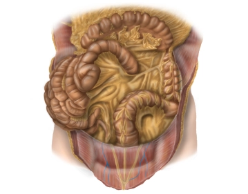
    The following parts of the intestine are relevant during the operation:

    • Descending colon:
      • Location: secondary retroperitoneal
      • Peritoneal suspension: fused with the posterior wall of the abdominal cavity
      • Course: from the left flexure (here: phrenocolic ligament with fixation to the spleen) to the iliac fossa, connects to the transverse colon and transitions into the sigmoid colon
      • Length: 20-30 cm
    • Sigmoid:
      • Location: intraperitoneal
      • Peritoneal suspension: sigmoid mesocolon
      • Course: from the iliac fossa as a loop (S-shaped) to the level of the 2nd-3rd sacral vertebra, connects to the descending colon and transitions into the rectum
      • Length: variable (elongated sigmoid), usually about 35 cm
    • Rectum:
      • Transition from sigmoid colon to rectum before the 2nd-3rd sacral vertebra.
      • Length 16 cm
      • Division into thirds: The height of the margins is measured with a rigid endoscope, using the anal-cutaneous line as a reference.
        • Lower third 0-6cm
        • Middle third 6-12cm
        • Upper third 12-16cm
        • each measured from the anal-cutaneous line
  2. Fascia systems

    • Fascia pelvis parietalis covers the pelvic wall with vessels, vegetative nerves, and presacral venous/nerve plexus.
    • Above the anorectal junction, the fascia rectosacralis meets the fascia pelvis visceralis.
    • The visceral fascia includes the fascia propria organi pelvis (surrounding the mesorectum dorsally and laterally) and the ventrally located Denonvilliers fascia.
    • The Denonvilliers fascia lines the posterior wall of the bladder, the seminal vesicles, and the posterior wall of the prostate in men.
  3. Vascular supply and lymphatic drainage

    Vascular supply and lymphatic drainage 1
    Vascular supply and lymphatic drainage 2
    Vascular supply and lymphatic drainage 3
    • Arterial supply of the left colon, sigmoid colon up to the upper rectum by the inferior mesenteric artery (A. mes. inf./AMI)
      • Origin of the A. mes. inf. from the abdominal aorta at the level of LWK 3
      • Left colic artery: supplies the descending colon, an ascending branch anastomoses with the middle colic artery, a descending branch anastomoses with a sigmoid artery
      • Sigmoid arteries: 2-4 arteries, several small branches to the sigmoid colon, anastomoses to the left colic artery and superior rectal artery
      • Superior rectal artery: runs from dorsal to the upper rectum, anastomoses with sigmoid artery and middle rectal artery from the internal iliac artery
      • Middle rectal arteries: from the internal iliac artery
      • Inferior rectal artery from the internal pudendal artery

    Cave: The inferior mesenteric plexus (inferior mesenteric ganglion) surrounds the origin of the A. mes. inf.

    • Special features:
      • Left colic flexure: watershed between the supply area of the superior mesenteric artery and the inferior mesenteric artery
      • Anastomosis between the superior mesenteric artery (middle colic artery) and the inferior mesenteric artery (left colic artery) near the left colic flexure distally (Riolan's anastomosis)
      • Drummond arcade: vascular arcade that connects the branches of the colic branches of the superior mesenteric artery and the inferior mesenteric artery near the colon and runs caudally.

    Cave: Inconstant: in 20% of cases, the Riolan anastomosis is not or not sufficiently developed.

    • Arterial supply of the rectum:
      • Upper third of the rectum via the superior rectal artery from the A. mes. inf., which divides into two terminal branches dorsally,
      • Middle third from the middle rectal artery (paired, each as a branch of the internal iliac artery) The middle rectal arteries run in the so-called lateral ligaments of the rectum and are severed during total mesorectal excision.
      • Lower third via the inferior rectal artery (paired, each as a branch of the internal pudendal artery from the internal iliac artery.


    • Venous drainage
      • Left hemicolon up to the upper rectum via the left colic vein, sigmoid veins, superior rectal veins into the inferior mesenteric vein, which drains into the splenic vein behind the tail of the pancreas. This forms the portal vein at the confluence of the portal veins with the superior mesenteric vein and other visceral veins behind the head of the pancreas
      • Lower rectum via the drainage area of the inferior vena cava.


    • Lymphatic drainage
      • For all sections of the rectum along the course of the superior rectal artery and the inferior mesenteric artery.
      • In the lower third additionally via lymphatic vessels along the middle rectal arteries and internal iliac arteries to lymph nodes in the area of the levator ani muscle and the ischiorectal fossa.
    • Lymph nodes individually:
      • The large group of inferior mesenteric lymph nodes forms its own group at the upper rectum (superior rectal lymph nodes),
      • The internal iliac lymph nodes take up lymph from the middle rectum (from the pararectal lymph nodes),
      • The superficial inguinal lymph nodes drain lymph from the lower anal region, the anus, and the skin of the perineal region.


    • Important neural structures:
      • Inferior mesenteric plexus (inferior mesenteric ganglion) (autonomic nervous system)
        • at the origin of the A. mes. inf.
        • Involved sympathetic nerves: lumbar splanchnic nerves, involved parasympathetic nerves: pelvic splanchnic nerves via the inferior hypogastric plexus, superior hypogastric nerves.
      • Superior hypogastric plexus
        • is located at the bifurcation of the aorta
        • Involved sympathetic nerves: fibers from the abdominal aortic plexus, lumbar splanchnic nerves
        • Involved parasympathetic fibers: pelvic splanchnic nerves (S2-S4)
        • Target organs: left hemicolon to rectum, bladder, ureter, prostate, uterus, vagina
      • Inferior hypogastric plexus
        • On both sides of the rectum to the bladder
        • Involved sympathetic fibers: sacral splanchnic nerves
        • Via hypogastric nerves connection to the superior hypogastric plexus
        • Involved parasympathetic fibers: pelvic splanchnic nerves (from S2–S4)
        • Target organs: descending colon, sigmoid, rectum, bladder, ureter, uterus, vagina, prostate
  4. Topographical relations

    Topographical relations 1
    Topographical relations 2
    • The descending colon runs from cranial to caudal adjacent to the left lateral abdominal wall. It has a close anatomical relationship to the:
      • spleen (flexure of the left colon),
      • omental bursa (dorsomedial of the flexure of the left colon),
      • left kidney
      • tail of the pancreas (cranial section of the descending colon).

    Cave: The flexure of the left colon is fixed to the diaphragm by the lig. phrenicocolicum and to the spleen by the lig. splenocolicum.

    • The sigmoid colon runs in an S-shape from the left iliac fossa into the small pelvis. The root of the sigmoid mesocolon crosses from the lateral iliac fossa medially:
      • the common iliac vessels,
      • the ureter
      • the ovarian or testicular vessels.
    • Rectum: Adjacent structures of the rectum are posteriorly the sacrum and coccyx, laterally next to the internal iliac artery and vein the regional lymph nodes, the sacral plexus, and parts of the autonomic nervous system as well as the ureter and adnexa. Ventrally, in the female organism, are the uterus and vagina, in the male the bladder and prostate/seminal vesicle. Caudally is the pelvic floor.

    Cave: Course of the ureter:

    • Dorsal of the descending colon and sigmoid colon under the Gerota fascia from the kidney radiating caudally
    • on the psoas muscle, which it crosses from laterocranial to caudomedial.
    • It drains from laterocranial lateral of the upper rectum into the bladder