Start your free 3-day trial — no credit card required, full access included

Anatomy - Total gastrectomy, robotically assisted with D2 lymphadenectomy

  1. Surgical Anatomy of the Stomach/Macroscopy

    506_Anatomie_Magen_01.jpeg
    • The stomach is a muscular hollow organ between the esophagus and duodenum. It is located in the left and middle upper abdomen directly below the diaphragm. The stomach is on average 25 to 30 cm long when moderately filled and has a capacity of 1.5 liters, in extreme cases up to 2.5 liters. The position, size, and shape of the stomach vary greatly depending on age, filling state, and body position. There are large interindividual differences.
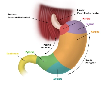
    • The stomach is divided into different sections:
      • Cardia (stomach entrance, upper stomach mouth, Ostium cardiacum):
        The cardia is a 1-2 cm long area where the esophagus opens into the stomach. Here is the sharp transition from the esophageal mucosa to the gastric mucosa, which is usually well recognizable with the endoscope.
      • Fundus gastricus: Above the stomach entrance, the fundus arches upwards, also called the stomach dome or fornix gastricus. The fundus is usually filled with air that is involuntarily swallowed while eating. In a standing person, the fundus forms the highest point of the stomach, so that in an X-ray image the accumulated air is visible as a "stomach bubble." Opposite the stomach entrance, the fundus is demarcated by a sharp fold (Incisura cardialis).
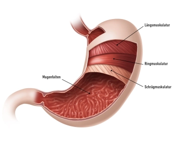
      • Corpus gastricum (stomach body): The stomach body forms the main part of the stomach. Here are deep longitudinal folds of the mucosa (Plicae gastricae), which extend from the stomach entrance to the pylorus and are also referred to as the "gastric canal."
      • Pylorus (Pars pylorica, stomach gatekeeper):
        This section begins with the expanded antrum pyloricum, followed by the gatekeeper canal (Canalis pyloricus) and ends with the actual stomach gatekeeper (Pylorus). Here is the stomach sphincter muscle (M. sphincter pylori), which consists of a strong circular muscle layer and closes the lower stomach mouth (Ostium pyloricum). The pylorus regulates the stomach exit and periodically allows small amounts of food pulp (chyme) to pass into the subsequent duodenum.

    The stomach is located intraperitoneally and is thus covered with a serosa layer; only the dorsal cardia is free of serosa. Due to the embryonic stomach rotation, the original mesogastria move from their sagittal position to a frontal orientation: The lesser omentum runs from the lesser curvature to the liver portal, while the greater omentum spreads from the greater curvature to the transverse colon, spleen, and diaphragm.

    The stomach is anchored and stabilized in the abdominal cavity by ligaments that extend to the liver and spleen, among others. With its convex side, it forms the greater curvature (Curvatura major) and with its concave side the lesser curvature (Curvatura minor). The front wall is referred to as Paries anterior and the back wall as Paries posterior. The greater omentum originates from the area of the greater curvature, while the lesser omentum spans between the left liver lobe and the lesser curvature.

    Topographical Anatomy of the Stomach

    Ventral (front):

    • The front wall of the stomach lies mostly directly against the abdominal wall.
    • In the upper area, it borders on the left liver lobe (Lobus sinister hepatis).

    Dorsal (back):

    • Behind the stomach lies the omental bursa (lesser sac).
    • Important neighboring structures are the pancreas (Pankreas), the spleen (Splen), the left kidney, and the adrenal gland.

    Cranial (above):

    • The stomach entrance (Cardia) is in direct proximity to the esophagus (Ösophagus) and the diaphragm.
    • The fundus is located below the left diaphragm area and borders on the spleen.

    Caudal (below):

    • The stomach transitions through the pylorus into the duodenum, which is already located in the right upper abdomen.

    Lateral (sideways):

    • On the left side, the stomach is bordered by the spleen, which is connected to it via the Lig. gastrosplenicum.
    • On the right side, the lesser curvature is in contact with the liver portal (Porta hepatis).
  2. Layers and structure of the stomach wall/microscopy

    506_Anatomie_Magen_02.jpeg

    Stomach Wall
    The stomach wall exhibits a characteristic layered structure from inside to outside under the microscope:

    Tunica mucosa:
    The Tunica mucosa is the mucosal layer lining the stomach from the inside. It is divided into three sublayers:

    • The Lamina epithelialis mucosae forms a viscous, neutral mucus that protects the gastric mucosa from mechanical, thermal, and enzymatic damage.
    • Beneath it lies the Lamina propria mucosae as a shifting layer in which the gastric glands (Glandulae gastricae) are embedded.
    • Finally, the Lamina muscularis mucosae, a thin muscle layer, follows, which can alter the relief of the mucosa.

    Tela submucosa:
    This loose shifting layer consists of connective tissue and contains a dense network of blood and lymph vessels as well as the Plexus submucosus (Meissner's Plexus), a nerve fiber network that controls gastric secretion. This plexus operates largely autonomously but is influenced by the autonomic nervous system.

    Tunica muscularis:
    The Tunica muscularis is a strong muscle layer consisting of three layers with differently oriented muscle fibers:

    • an inner layer with obliquely oriented muscle fibers (Fibrae obliquae),
    • a middle circular muscle layer (Stratum circulare),
    • an outer longitudinal muscle layer (Stratum longitudinale).
      These muscle layers enable the peristalsis of the stomach and ensure the mixing of the chyme with gastric juice. Between the circular and longitudinal muscle layers runs the Plexus myentericus (Auerbach's Plexus), which controls muscle movements. Like the Plexus submucosus, this plexus operates largely autonomously but is influenced by the autonomic nervous system.

    Tela subserosa and Tunica serosa:
    Following another connective tissue shifting layer (Tela subserosa) is the Tunica serosa, which is divided into several layers:

    • The Lamina propria serosae contains blood and lymph vessels, nerves, and immune defense cells, referred to as milk spots (Maculae lacteae).
    • The Lamina epithelialis serosae, directed towards the body cavity, consists of a single-layered squamous epithelium (Serosaepithel). This shiny, transparent layer ensures good mobility of the stomach against adjacent organs via a thin film of fluid.

    Gastric Glands
    The gastric glands (Glandulae gastricae) are located in the Lamina propria mucosae and are mainly found in the fundus and corpus of the stomach. Up to 100 glands are located on 1 mm² of the mucosal surface. Various cell types are found in the glandular tubes:

    Mucous Cells:
    These cells produce the same neutral mucus as the epithelial cells, thus contributing to the protection of the gastric mucosa.

    Neck Cells:
    These superficially located cells secrete alkaline mucus, which has a high pH value due to the contained bicarbonate ions (HCO₃⁻). This mucus regulates the pH value of the stomach and protects the mucosa from self-digestion by hydrochloric acid (HCl) and enzymes. These cells are particularly numerous in the cardia and fundus of the stomach.

    Chief Cells:
    Chief cells produce the inactive precursor enzyme Pepsinogen. This is converted into the active enzyme Pepsin by hydrochloric acid (HCl), which digests dietary proteins. Since activation occurs only at the surface of the gland, self-digestion of the glands is prevented. Chief cells are mainly found in the corpus of the stomach.

    Parietal Cells:
    These cells, which are frequently found in the gastric corpus, produce hydrogen ions (H⁺) needed for the production of hydrochloric acid (HCl). Hydrochloric acid has a very low pH value of 0.9–1.5. Additionally, parietal cells produce the so-called Intrinsic Factor, which forms a complex with vitamin B12 in the intestine, enabling the absorption of the vitamin. Vitamin B12 is essential for erythropoiesis. A deficiency due to stomach removal can therefore lead to anemia.

    G Cells:
    These cells are preferably located in the antrum of the stomach and produce the hormone Gastrin, which stimulates the production of hydrochloric acid in the parietal cells

  3. Function

    The stomach serves as a reservoir for ingested food and plays a central role in digestion. Its main functions are the storage, mixing, and preliminary digestion of food. The food bolus (Chymus) is gradually released in controlled portions through the pylorus (Pylorus) into the duodenum, where the actual digestion continues.

    Storage and Mixing of Food

    The stomach can accommodate large amounts of food, which are stored over several hours. This allows for a constant energy supply and makes it possible to meet daily nutritional needs with just a few larger meals. The food is mechanically broken down in the stomach by the strong muscles of the Tunica muscularis and mixed with gastric juice through rhythmic contractions until a homogeneous chyme is formed.

    Formation and Function of Gastric Juice

    Gastric juice is continuously produced in the stomach, consisting of mucus, hydrochloric acid (HCl), enzymes, and other components:

    • Mucus (Mucin): The mucus produced by epithelial and neck cells protects the gastric mucosa from the aggressive effects of hydrochloric acid and mechanical damage.
    • Hydrochloric Acid (HCl): It is produced by the parietal cells (Parietalzellen) and creates a highly acidic environment (pH 0.9–1.5). This acid has several important functions:
      • It denatures food proteins, preparing them for enzymatic digestion.
      • It activates the inactive pepsinogen, produced by the chief cells, into the active form Pepsin, which breaks down proteins into smaller peptides.
      • It acts as an antibacterial agent, preventing pathogens from entering the intestines through food.

    Preliminary Digestion of Food Components

    The stomach initiates the digestion of proteins. The enzyme Pepsin cleaves larger proteins into smaller polypeptides, preparing the food for further processing in the small intestine. Carbohydrates and fats are only partially digested in the stomach, as the main work of their digestion occurs in the small intestine.

    Regulation of Stomach Function

    The stomach function is regulated by a complex interplay of nerves and hormones:

    • The Plexus myentericus and the Plexus submucosus control stomach movements and gastric juice secretion. Both operate autonomously but are influenced by the autonomic nervous system (vagal and sympathetic).
    • Hormones such as Gastrin, produced by the G-cells in the antrum of the stomach, promote the production of gastric juice and stimulate the contractions of the stomach muscles.

    Transfer of the Chymus

    After the storage and processing of food, the chyme is transported towards the pylorus by the peristaltic movements of the stomach muscles. The Pylorus opens only temporarily to release small amounts of the chymus into the duodenum. This ensures that the small intestine is not overloaded and that digestive enzymes and bile salts can work effectively.


    Interaction of the Stomach with Other Organs

    The stomach is centrally involved in digestion and works closely with various organs to optimally prepare nutrients from food:

    • Esophagus: The stomach receives food through the lower esophageal sphincter (Cardia), which prevents the backflow of gastric juice into the esophagus.
    • Liver and Gallbladder: The liver produces bile, which is stored in the gallbladder. After passing through the stomach, the chymus enters the duodenum, where bile is released to emulsify fats.
    • Pancreas: The pancreas produces digestive enzymes (e.g., amylase, lipase, proteases) and bicarbonate, which neutralizes the acidic food bolus. These enzymes are also released into the duodenum.
    • Small Intestine: The stomach releases the pre-digested chymus in small portions into the duodenum, where the main digestion and absorption of nutrients occur. The duodenum provides feedback to the stomach via hormones such as Secretin and Cholecystokinin to regulate gastric acid secretion and emptying.
    • Spleen: Although the spleen is not directly involved in digestion, it indirectly supplies the stomach with blood via the A. splenica. It also plays an important role in immune defense and filters microorganisms that may enter the body through food.

    This complex interaction ensures that the chyme is gradually processed and prepared for absorption in the intestine. Hormones, nerves, and local reflexes coordinate the processes between the organs and adapt them to the respective food situation.

  4. Arterial, venous, and neural supply

    506_Anatomie_Arterien besch.jpg
    506_Anatomie_venen besch.jpg

    Arterial Supply

    The arterial supply of the stomach is provided by several blood vessels, all originating from the unpaired Truncus coeliacus. These vessels run along the two curvatures of the stomach and form a network of vascular arcades that comprehensively supply the stomach. The main arteries are:

    • Right gastric artery: This arises from the proper hepatic artery and runs along the lower part of the lesser curvature.
    • Left gastric artery: It supplies the upper part of the lesser curvature and is a direct branch of the Truncus coeliacus.
    • Short gastric arteries: These small arteries arise from the splenic artery and supply the fundus of the stomach.
    • Right gastroepiploic (gastro-omental) artery: It arises from the gastroduodenal artery and runs along the right part of the greater curvature.
    • Left gastroepiploic (gastro-omental) artery: This also arises from the splenic artery and supplies the left part of the greater curvature.
    • Posterior gastric artery: This artery, a branch of the splenic artery, supplies the posterior wall of the stomach.

    The supply of the stomach is mainly through two vascular arcades:

    1. Lesser curvature: Here, the left gastric artery and right gastric artery form an anastomosis.
    2. Greater curvature: Here, the supply is through the left gastroepiploic artery and right gastroepiploic artery, which also anastomose with each other.

    Venous Supply

    The venous drainage of the stomach parallels the arterial supply. Four large veins run along the two curvatures of the stomach and eventually drain the blood through the portal vein of the liver:

    • Left and right gastric veins: These veins accompany the arteries of the same name along the lesser curvature and drain directly into the portal vein of the liver.
    • Left gastroomental (gastroepiploic) vein: This vein runs along the left part of the greater curvature and drains into the splenic vein.
    • Short gastric veins: They accompany the short gastric arteries and also drain into the splenic vein.
    • Right gastroomental (gastroepiploic) vein: This vein runs along the right part of the greater curvature and drains into the superior mesenteric vein.

    All collecting veins ultimately drain into the portal vein (portal vein), allowing the venous blood of the stomach to reach the liver.

    Nervous Supply

    The nervous supply of the stomach is provided by the autonomic nervous system and includes both sympathetic and parasympathetic components. Additionally, sensory nerve fibers are involved.

    Sympathetic:
    The sympathetic component innervates particularly the musculature of the pylorus (Pylorus). The sympathetic nerve fibers originate from the celiac ganglion.

    Parasympathetic (Vagus nerve):
    The vagus nerve (10th cranial nerve) supplies the rest of the stomach musculature as well as the glands of the stomach.

    • The right vagus branch forms the posterior vagal trunk and runs to the posterior wall of the stomach.
    • The left vagus branch forms the anterior vagal trunk and innervates the anterior wall of the stomach.
      Both vagus branches enter the abdominal cavity through the esophageal hiatus of the diaphragm.

    Sensory fibers:
    The afferent sensory fibers of the stomach travel via the greater splanchnic nerve and terminate in the thoracic spinal ganglia. These fibers are responsible for the perception of pain and distension of the stomach.

  5. Lymphatic drainage pathways

    506_Anatomie_Lymphe besch2 Kopie.jpg

    Lymphatic Drainage of the Stomach

    The lymphatic vessels of the stomach run parallel to the arterio-venous supply and drain into various lymph node stations. This drainage plays a crucial role in the spread of tumor cells and the diagnosis of gastric carcinomas.

    Lymphatic drainage by regions:

    • Lesser curvature:
      The lymph flows parallel to the Aa. gastricae sinistra et dextra and is drained into the Nll. gastrici sinistri et dextri.
    • Fundus:
      The lymph from the fundus flows along the A. splenica into the Nll. splenici.
    • Greater curvature:
      The lymph follows the suspension of the Omentum majus (greater omentum) and drains into the Nll. gastroomentales dextri et sinistri.
    • Pyloric region:
      The lymph from the pyloric area is drained into the Nll. pylorici.

    Further lymph flow:
    From the regional lymph nodes, the lymph flows further:

    • Into the Nll. coeliaci (lymph nodes at the celiac trunk).
    • To the superior mesenteric lymph nodes.
    • Via the thoracic duct into the venous system.

    Special feature – Virchow's node:
    An advanced gastric carcinoma can lead to metastasis in the left neck region, where the so-called Virchow's node (in the left supraclavicular fossa) becomes noticeable. This finding is an important clinical sign of disseminated tumor spread.

    Lymph Node Stations by Compartments

    For surgical reasons, the lymph node stations of the stomach are divided into three compartments:

    Compartment I (LN group 1–6):
    Lymph nodes directly at the stomach:

    • Paracardial (Group 1 and 2).
    • At the lesser and greater curvature (Group 3 and 4).
    • Suprapyloric and infrapyloric (Group 5 and 6).

    Compartment II (LN group 7–11):
    Lymph nodes along the major vessels:

    • A. gastrica sinistra (Group 7).
    • A. hepatica communis (Group 8).
    • Truncus coeliacus (Group 9).
    • Splenic hilum (Group 10).
    • A. lienalis (Group 11).

    Compartment III (LN group 12–16):
    Lymph nodes in the vicinity of the stomach and major abdominal vessels:

    • Lig. hepatoduodenale (Group 12).
    • Behind the pancreatic head (Group 13).
    • At the mesenteric root and mesentery (Group 14 and 15).
    • Along the abdominal aorta (Group 16).

    Clinical Relevance

    The detailed knowledge of the lymphatic drainage and lymph node stations of the stomach is crucial, especially in the planning of surgeries (mandatory oncological lymphadenectomy in gastric carcinomas). The lymph node compartments help to define the resection boundaries and to determine the tumor stage postoperatively with precision.