
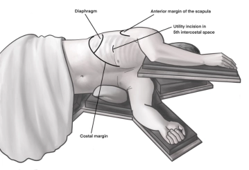
Incision of approximately 4 cm in length in the area of the anterior axillary line at the upper edge of the 5th rib to access the 4th intercostal space above. A helpful orientation is often an imaginary line from the tip of the scapula to the nipple. Transection of the subcutis on the rib with the monopolar knife. Subsequently, stepwise preparation of the intercostal muscles with the monopolar knife. The pleura is opened bluntly with a finger. Palpation of the thoracic wall for adhesions and insertion of a wound protection film.
-
Access uniportal VATS right
![Access uniportal VATS right]()
-
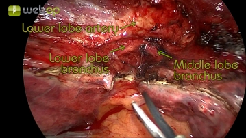
Exploration of the thorax and preparation of the pulmonary artery in the lobar fissure
![Exploration of the thorax and preparation of the pulmonary artery in the lobar fissure]()
Soundsettings Initially, the exploration of the thorax is performed for macroscopically suspicious lesions and adhesions. Subsequently, the preparation begins at the interlobium. Due to the well-developed fissure, the interlobar part of the pulmonary artery with the underlying bronchial system is clearly visible after incision of the visceral pleura. Then, the lymph nodes of station 11 (according to IALSC) are removed.
-
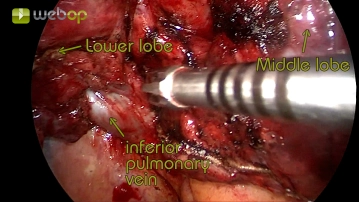
Preparation of the pulmonary ligament and visualization of the inferior pulmonary vein
![Preparation of the pulmonary ligament and visualization of the inferior pulmonary vein]()
Soundsettings After displaying the pulmonary artery, the pulmonary ligament is now prepared. Simultaneously, the lymph nodes of station 9 (according to IALSC) are removed. The preparation concludes with the display of the inferior pulmonary vein.
-
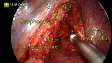
En-bloc resection of the lymph nodes of station 7 (according to IALSC)
![En-bloc resection of the lymph nodes of station 7 (according to IALSC)]()
Soundsettings Continue the dissection cranially with incision of the pleural hilum at the medial lung hilum. Here, the lymph nodes of station 8 (according to IALSC) and, after precise dissection, the lymph nodes of station 7 (according to IALSC) are removed as an en-bloc resection.
Preparation of the inferior pulmonary vein
By mostly blunt dissection of the lung parenchyma and situational use of the ultrasonic shears for
By mostly blunt dissection of the lung parenchyma and situational use of the ultrasonic shears for
Activate now and continue learning straight away.
Single Access
Activation of this course for 3 days.
US$9.10
inclusive VAT
Most popular offer
webop - Savings Flex
Combine our learning modules flexibly and save up to 50%.
from US$4.23 / module
US$50.80/ yearly payment
thoracic
Unlock all courses in this module.
US$8.46
/ month
US$101.60 / yearly payment
Webop is committed to education. That's why we offer all our content at a fair student rate.