The thoracic cage protects the internal organs and simultaneously allows respiratory excursion through its structure composed of bony, cartilaginous, and muscular parts. The thoracic spine and the sternum are movably connected by 10 pairs of ribs. Two additional pairs of ribs originate only from the thoracic vertebrae.
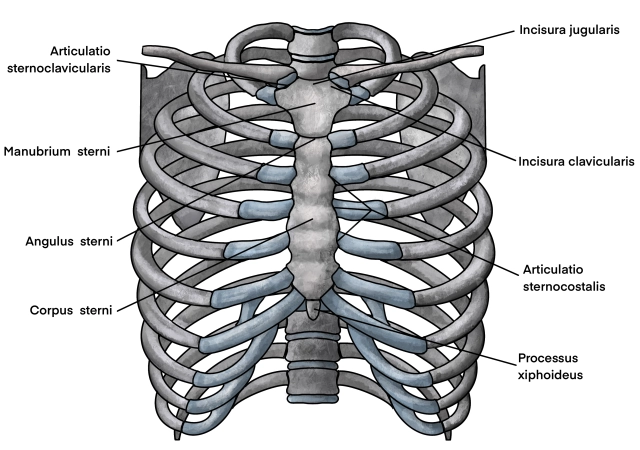
The sternum itself consists from cranial to caudal of three parts: the manubrium sterni, the corpus sterni, and the processus xiphoideus. These are connected by synchondroses, which ossify in adulthood.
The ventral ends of ribs 1 to 10 are connected to the costal cartilage via the costochondral joints. The costal cartilages of ribs 1 to 6 reach the sternum directly via the sternocostal joints, while those of ribs 7 to 10 form the costal arch through the interchondral joints and terminate at the cartilage of the 6th rib. The sternocostal joints are mainly reinforced by the radiate sternocostal ligaments, which form the membrana sterni ventrally on the sternum with its periosteum.
Between the ribs are the external and internal intercostal muscles, which laterally close the thoracic cage muscularly.
Internally, the thorax is lined by the endothoracic fascia and the parietal pleura.
It is important to note the internal thoracic vessels, which run somewhat lateral to the sternum parallel to it in a caudal direction, as well as the intercostal vessels and nerves, which are found at the lower edge of each rib.