Start your free 3-day trial β€” no credit card required, full access included

EVAR – Endovascular Repair of an Abdominal Aortic Aneurysm (Bifurcated Graft)

Reading time readingtime 33:44 min.
Activate now
  1. Exposing the femoral bifurcation, right groin

    Video
    Exposing the femoral bifurcation, right groin
    Soundsettings

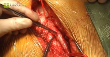
    Approximately 1 cm lateral to the palpable femoral artery, make a longitudinal skin incision on the proximal thigh distal to the inguinal region. After transecting the subcutaneous tissue, incise the femoral fascia longitudinally medial to the sartorius muscle. Open the fascia of the adductor canal at the caudal edge of the incision, expose the superficial femoral artery, and encircle it with a vessel loop. Divide and suture-ligate between Overholt forceps the lymph collectors crossing beneath the inguinal ligament. Encircle the common femoral artery and the deep femoral artery.

    Repeat the same manoeuvre to expose the femoral bifurcation in the left groin.

    Tips:

    1. Access lateral to the femoral artery spares the lymph collectors and permits subsequent offset layered closure to promote reliable wound healing.

    2. The superficial femoral artery serves as a landmark for dissecting proximally lateral to it. This allows the deep femoral artery and the common femoral artery to be located easily and also avoids the risk of injury to the veins running medially.

    3. Suture ligation of the lymph collectors helps, to a large extent, to prevent subsequent lymphatic cysts and fistulae. Postoperative lymph collection in the groin not infrequently paves the way for deep wound infection with fatal consequences.

    4. Circumflex femoral artery: at times, this artery lies hidden and can cause profuse back-bleeding if it is not clamped off.
  2. Puncturing the left common femoral artery, introducing a 6F sheath in Seldinger technique and placing a guidewire

    Video
    Puncturing the left common femoral artery, introducing a 6F sheath in Seldinger technique and placing a guidewire
    Soundsettings

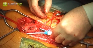
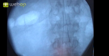
    The left common femoral artery is punctured with an 18G puncture needle and a 6F sheath is introduced using the Seldinger technique. 5000 IU of heparin-saline solution is administered locally into the vessel. Under fluoroscopic control, a guidewire with hydrophilic coating (here: Terumo®) is introduced into the descending aorta.

    Tip:

    In case of marked coiling, steering with a curved guiding catheter may be necessary.

  3. Puncturing the right common femoral artery, introducing a 6F sheath in Seldinger technique and placing a guidewire

    Video
    Puncturing the right common femoral artery, introducing a 6F sheath in Seldinger technique and placing a guidewire
    Soundsettings

    The same manoeuvre as in surgical step 2 for the right groin.

  4. Inserting the pigtail catheter on the left, central marking / renal artery level

    Video
    Inserting the pigtail catheter on the left, central marking / renal artery level
    Soundsettings

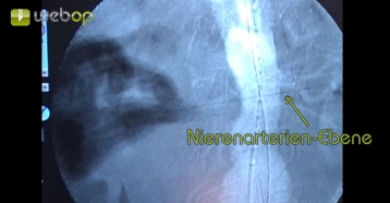
    A graduated pigtail catheter is now introduced via the indwelling guidewire on the left. Using angiography with a high-pressure injector, the renal artery level is identified and marked extracorporeally with a needle for rough orientation.

Peripheral marking / origin of both internal iliac arteries

After repeat injection of contrast medium, the origins of both internal iliac arteries are marked e

Activate now and continue learning straight away.

Single Access

Activation of this course for 3 days.

US$9.30  inclusive VAT

Most popular offer

webop - Savings Flex

Combine our learning modules flexibly and save up to 50%.

from US$4.30 / module

US$51.61/ yearly payment

price overview

vascular surgery

Unlock all courses in this module.

US$8.60 / month

US$103.20 / yearly payment

to top