Start your free 3-day trial — no credit card required, full access included

Perioperative management - Iliacomesenteric venous bypass in superior mesenteric artery occlusion—Vascular Surgery

  1. Indications

    • Intestinal angina due to short segmental stenosis of the superior mesenteric artery (SMA) close to its origin or occlusion due to inadequate collateral blood supply via the celiac trunk
    • Lack of indication for interventional treatment, e.g., in long stenosis at the origin stenosis/occlusion of the SMA).
    • Failure of interventional management

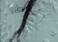
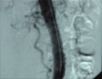
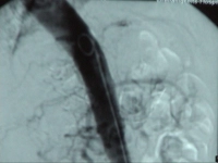
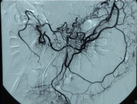
    Video clip: SMA occlusion close to its origin with occlusion of the celiac trunk

    PM 310-1
    DSA of the abdominal aorta in the anteroposterior plane: Occlusion of the inferior mesenteric artery and celiac trunk


    PM 310-2
    DSA of the abdominal aorta in the lateral plane: Occlusion of the inferior mesenteric artery and celiac trunk


    PM 310-3
    DSA of the abdominal aorta in the lateral plane: Occlusion of the inferior mesenteric artery and celiac trunk; late filling of the celiac trunk via collaterals


    PM 310-4
    DSA of the abdominal aorta in the anteroposterior plane: Probing of the inferior mesenteric artery (IMA) and visualization of the inferior mesenteric artery via the IMA.


    Grade of ischemia and general indication for surgery

    StageSymptomsIndication for surgery
    IAsymptomaticOptional in concurrent procedures for vascular occlusion, aortic or iliac artery interventions
    IIIntestinal angina = crampy postprandial abdominal pain, cachexiaAbsolute indication for surgery
    IIIAbdominal pain at restAbsolute indication for surgery
    IVAcute abdomen secondary to mesenteric infarction, intestinal gangrene, possibly peritonitis due to transmural bacterial translocationEmergent indication for surgery
  2. Contraindications

    • long-distance and multifocal stenoses of the SMA > 6 cm usualy do not lend themselves to open repair
    • Multisegment occlusive processes of the SMA, e.g. in thrombangitis obliterans (Winiwarter-Buerger disease) or lupus erythematosus
    • ASA IV
    • COPD Cold IV
  3. Preoperative diagnostic work-up

    Key aspects first:

    1. Chronic intestinal ischemia syndromes: Only diagnostic imaging yields useful insights!

    2. Usually there are multiple (vascular) comorbidities -> detailed examination by vascular surgeons and angiologists.

    3. Patient should undergo work-up to identify other abdominal diseases, especially to rule out malignancies.

    4. Delayed diagnosis > 12 months because differential diagnosis included the disease too late.

    Medical history

    Typical triad in malperfusion of the SMA:

    • postprandial abdominal pain starting shortly (about 20 min) after food intake and subsiding after 3-4 hours (intestinal angina)
    • reactive restriction on food intake -> weight loss, muscle atrophy, asthenia ("small-meal-syndrome")

    Absence of weight loss does not rule out the diagnosis!

    Ischemia of the celiac trunk distribution site results in nausea, vomiting, and meteorism; constipation is more likely to result from inferior mesenteric artery disease.

    Duplex ultrasonography for assessment of the intestinal vessels

    • Stenosis typically at the origin
    • Increased flow rate—flow rates greater than 200 cm/s are considered pathognomonic for high-grade stenosis—as well as turbulent flow

    CT angiography or MR angiography

    • with image reconstruction

    digital subtraction angiography of the intestinal arteries in 2 planes

    • when planning an intervention

    Abdominal ultrasonography

    (Exercise) ECG

    Chest X-ray

    Laboratory panels

    • Blood count, electrolytes, coagulation, renal retention parameters, blood lipids

    Staged diagnostic approach to intestinal ischemia


    Stage I:

    Stage II

    Stage III

    Stage IV

    Medical history and clinical examination

    +

    +

    +

    +

    Duplex ultrasonography

    +

    +

    +

    Ø

    angiography

    Ø

    +

    +

    +

    CT/MR angiography

    Ø

    +

    +

    Ø

  4. Special preparation

    • Ultrasound assessment of whether an autologous great saphenous vein is available as a bypass (possibly synthetic graft)
    • Identify blood group, provide for packed RBCs, if needed
    • Enema
    • Indwelling urinary catheter
  5. Informed consent

    General surgical risks

    • Major bleeding, blood transfusions, transmission of hepatitis/HIV through allogeneic blood transfusions
    • Allergy/intolerance
    • Wound infection
    • Thrombosis/embolism
    • Skin/vascular/nerve damage, e.g., due to patient positioning
    • Keloid

    Risks special to this procedure

    • Incisional hernia
    • Abdominal adhesions, ileus
    • intraoperative peripheral embolism in iliac/leg arteries -> surgical intervention, in case of ischemic leg including amputation
    • Massive infection → bleeding, peritonitis, sepsis; possible reoperation with explantation of the vascular graft
    • Injury to neighboring organs (ureter, bladder, bowel, liver, spleen); intestinal stoma
    • Impaired perfusion or nerve injury of the spinal cord (paresis, erectile dysfunction)
    • In case of autologous vein bypass: Secondary bleeding, infection, keloid
    • Intraoperative angiography: Contrast agent → renal failure
    • Bypass infection/stenosis → impaired perfusion of the affected organs, reoperation
    • Impaired intestinal perfusion → defecation disorder, malabsorption, ischemic colitis → resection, possibly colostomy
  6. Anesthesia

  7. Positioning

    Positioning

    Supine position, right arm adducted, left arm abducted. The upper body should be slightly retroflexed at the level of the thoracoabdominal transition between the pubic bone and xyphoid to increase the distance between the iliac crest and costal arch.

  8. Operating room setup

    Operating room setup

    Rechts des Patienten steht der Operateur, ihm gegenüber die Assistenz. Die instrumentierende OP-Fachkraft steht fußwärts neben der Assistenz.

  9. Special instruments and fixation systems

    • Abdominal wall retractor
    • Aortic clamps, bulldog clamps
    • Monofilament, non-absorbable vascular sutures (3/0 to 6/0)
    • Cell-Saver
  10. Postoperative management

    Postoperative analgesia:

    Follow this link to PROSPECT (Procedures Specific Postoperative Pain Management) and to the current German guideline Behandlung akuter perioperativer and posttraumatischer Schmerzen [Treatment of acute perioperative and posttraumatic pain].

    Postoperative care

    • ICU for 24 hours, then IMC
    • Cardiovascular stabilization, monitoring of pulmonary function
    • Balanced infusion and transfusion management
    • Broad-spectrum antibiotics
    • Laboratory monitoring (serum lactate, leukocytes, CRP, acid-base status)
    • Monitoring of leg pulses, assessment of capillary perfusion in the feet.
    • Early return to enteral nutrition

    Anticoagulation

    Heparin

    • Postoperative day 1: 7500 IU via infusion pump
    • Postoperative day 2–4: 15000 IU via infusion pump
    • Starting on postoperative day 5: low molecular weight heparin

    Phenprocoumon for at least 6 months or even lifelong

    • In repair with long synthetic grafts or venous bypasses, e.g., iliacoceliac, iliacomesenteric
    • In multiplevascular occlusions

    ASA 100 mg

    • Direct repair
    • Short bypasses, e.g., aorto-celiac (subdiaphragmatic)
    • Interposition grafts, aortoceliac/-mesenteric or aortoarterial

    Ambulation

    • Starting on postoperative day 2 sitting on edge of bed

    Physical therapy

    • Isometric exercises
    • Breathing exercises

    Diet

    • Early return to enteral nutrition, e.g. with high-calorie oral formula
    • Possibly weight gain by eating several meals a day, rich in carbohydrates, no foods causing flatulence

    Bowel movement

    • Possibly on postoperative day 3 (enema, amidotrizoate acid; neostigmine as last resort)

    Work disability

    • Around 3 months

    Always remember!

    • Risk reduction (treatment of the underlying disease, no mnore nicotine, lowering of cholesterol levels)
    • Annual or biennial follow-up with duplex ultrasonography