The diagnosis of acute limb ischaemia can often be made "at a glance" after a brief history and examination. As a rule, angiography is not performed (exception: concurrent PAOD) and management is planned based on the clinical findings (history, inspection, palpation). After the absolute ischaemia has been resolved, diagnosis and treatment of the source of the embolism can be approached in an orderly fashion.
Emergent diagnostic work-up should address the following questions:
- Severity of ischaemia (Pratt's 6 Ps, TASC in PAOD)?
- Localisation and extent of the occlusion?
- Acute ischaemia resulting from embolisation or on the basis of PAOD?
The typical 6 Ps of Pratt characterise the severe course of acute limb ischaemia with a grave threat to limb viability (Source: Pratt GH (1954) Cardiovascular surgery. Kimpton, London):
In PAOD, the classification of acute limb ischaemia according to the Transatlantic Inter-Society Consensus (TASC) Working Group seeks to reflect the varying levels of urgency for further diagnostic work-up and management:
Medical history/clinical picture
Rough distinction between acute thrombotic occlusion and embolism:
→ Embolism
- Sudden, occasionally whip-like pain in the limb (cardinal symptom)
- Often severe ischaemia
- Cardiovascular history: known atrial fibrillation; heart defect; history of myocardial infarction; aortic aneurysm
→ thrombotic occlusion
- More likely symptoms increasing over several days, typically with incomplete ischaemia
- Known PAOD (claudication symptoms?)
- History of bypass surgery, stenting/PTA
- Atherosclerosis risk profile
- Local trauma
- No evidence of a source of the embolism
Skin colour
- White ischaemia: pale distal to the occlusion
- Blue ischaemia: prognostically less favourable, because thrombus formation due to stasis has already spread to the venous circulation
- The spontaneous Ratschow test may be seminal in incomplete ischaemia (total pallor with the limb elevated that does not recover even after the limb is subsequently returned to the horizontal position).
Capillary refill time
- brief plantar pressure with the finger on the big toe or forefoot → the initially pale pressure point normally turns red within < 3 seconds
- the longer the redness takes to appear, the more marked the perfusion disorder
Effective perfusion pressure
- Elevation of the limb allows the perfusion pressure to be estimated → 10 cm = 7.5 mmHg
Skin temperature
- The affected limb is colder on bilateral comparison
- The occlusion lies markedly more proximal than the drop in temperature
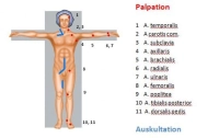
Pulses
- In the simplest case, absence of pulses in the affected limb
- Bilateral absence of pulses: pre-existing bilateral PAOD or asymptomatic embolism
Auscultatory bilateral comparison of the limb arteries
Ankle-Brachial Index (ABI)
- ABI = systolic BP of the posterior tibial artery / systolic BP of the brachial artery
- the lower the ABI, the more pronounced the ischaemia
- In the acute stage, the pressure may not be measurable
Colour-flow Doppler ultrasonography
- Carotid artery, abdominal aorta, limb arteries (rule out popliteal aneurysm in lower leg occlusions!)
- Localisation of stenoses and occlusions in almost all vascular regions except the thoracic area
- Quantification of the degree of stenosis and assessment of plaque morphology possible
- Sensitivity and specificity around 90%
CT angiography
- Multislice computed tomography (MS-CT) using a non-ionic contrast agent
- Broad range of indications: traumatic vascular lesion (esp. trunk); vascular dissection/rupture; aneurysm; arterial thrombosis/embolism; portal vein/mesenteric vein thrombosis; pulmonary artery embolism; PAOD; vascular tumours
- Advantages: rapidly performed; detects relevant comorbidities; visualises peripheral arteries; sensitivity and specificity each about 90%
- Disadvantages: radiation and contrast agent exposure, allergies (about 3%), no functional assessment
- The indication does not depend on the degree of ischaemia, but on the history and clinical findings in the contralateral leg: in case of pre-existing PAOD or evidence of a popliteal aneurysm, angiography is preferable. The findings then determine the surgical strategy. If PAOD or a popliteal aneurysm is concurrently present as the cause of the acute ischaemia, more complex reconstructions become necessary (e.g. interposition grafts, bypass procedures). However, in case of complete ischaemia too much time should not be lost on angiography (suitable logistics).
Laboratory panels
- Blood count
- Electrolytes
- Coagulation
- Kidney function parameters
- Liver enzymes
- Blood lipids
- Blood group
Cardiac check
Chest x-ray
As an immediate measure
administer 5000-10000 IU heparin to prevent appositional thrombosis. Wrap the foot in a cotton-wool bootie to prevent pressure injury (particularly intraoperatively as well!).