Right femoropopliteal PTFE bypass (P3) – Vascular Surgery - vascular surgery
You have not purchased a license - paywall is active: to the product selection
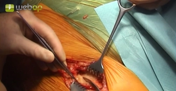
Exposing the femoral bifurcation, right groin
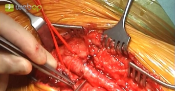
Approximately 1 cm lateral to the palpated femoral artery make a longitudinal skin incision on the proximal thigh distal to the inguinal aspect. After transecting the subcutaneous tissue, divide the femoral fascia longitudinally medial to the sartorius muscle. Open the fascia of the adductor canal at the distal edge of the incision, expose the superficial femoral artery, and encircle it with a vessel loop. Divide and suture ligate between Overholt forceps the lymph collectors crossing posterior to the inguinal ligament. The lymph nodes are enlarged because of PAOD Fontaine grade IV.
Tips:
1. Access lateral to the femoral artery spares the lymph collectors and permits subsequent offset layered closure to promote effective wound healing.
2. The superficial femoral artery serves as a landmark for proximad dissection lateral to it. This helps to easily locate the deep and common femoral arteries and also avoids the risk of injury to the veins running medially.
3. Suture ligation of the lymph collectors helps, to a large extent, to prevent subsequent lymphatic cysts and fistulae. Postoperative inguinal lymph collection in PAOD grade IV often paves the way for deep wound infection with fatal consequences for the limb and possibly even for the patient's life.
Dissecting the femoral bifurcation
Suprapopliteal exposure of popliteal artery segment P1
Access the first popliteal artrey segment through a medial thigh skin incision above the knee (transition from the middle to the distal third of the depth of the thigh). After transecting the subcutaneous tissue, divide the femoral fascia anterior or ventral to the sartorius muscle longitudinally and retract the muscle posteriad. Insert a retractor (e.g., from van Dongen retractor). Identify the NV bundle iat the inferior margin of the adductor canal, transect the popliteal fat pad and expose the first segment of the popliteal artery. Divide any crossing veins during dissection between ligatures. Clamp the popliteal artery after encircling it.
Attempted thrombectomy of popliteal artery segment P1
Contrary to the preoperative angiogram, the severely stenosed arteriosclerotic popliteal artery is thrombosed in segments 1 to 3 The thrombus is already starting to become organized and to adhere to the wall. Therefore, the attempted thrombectomy with the Fogarty catheter via the longitudinally incision of the popliteal artery is unsuccessful. The popliteal artery is ligated proximal and distal to the arteriotomy (not shown in the video).
Infrapopliteal exposure of popliteal artery segment P3
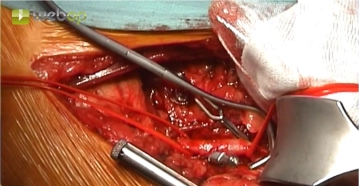
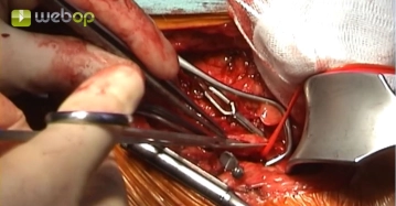
Incise the skin below the knee medial to the edge of the tibia. Coagulate any smaller vessels while dividing the subutaneous tissue. Divide the firm crural fascia longitudinally approximately 1 cm from the edge of the tibia. This exposes the medial head of the gastrocnemius muscle, which can be bluntly retracted posteriad. Insert a retractor between the muscle and posterior edge of the tibia. Expose the popliteal artery in the popliteal adipose tissue. Encircle the artery, clamp and incise it longitudinally. After checking inflow and back flow, instill 2000 IU heparin saline solution proximad and distad.
Tips:
1. During infrapopliteal exposure of the vessels, the femur can be rested on rolled up drapes within a sterile covering. Care must be taken to ensure that the knee joint and lower leg are exposed in slight flexion and not compressed. However, the correct length of the bypass must be measured with the leg fully extended because otherwise the bypass will be too short.
2. If a venous bypass is planned, care must be taken during skin incision and dissection through the subcutaneous tissue to avoid injury to the great saphenous vein as this may be a possible bypass graft. In such cases, it should be retracted posteriad for protection.
Preparing the PTFE graft
Fashioning the peripheral side-to-end anastomosis and tunneling the graft from segments P1 to P3
Pulling through the graft from segment P2 to the groin
TEA of femoral bifurcation and deep femoral artery
Reparing the vgraft, commencing the proximal side-to-end anastomosis
Completing the proximal side-to-end anastomosis and intraoperative angiography
Sealing the graft puncture sitewith a fascial patch
Single Access
Access to this lecture
for 3 days
€4.99 inclusive VAT

webop-Account Single
full access to all lectures
price per month
for the modul: General Surgery