Dialysis access surgery: Brachiocephalic fistula (“direct antecubital fistula”) - Vascular Surgery

Reading time readingtime 16:47 min.
Activate now
  1. Access in the right cubital fossa

    Video
    Access in the right cubital fossa

    Oblique skin incision in the right anticubital fossa with transection of the subcutaneous tissue.

    Tip:

    Spare the basilic and median cubital veins. They may come into play if the further course of the cephalic vein reveals problems and is unsuitable for fashioning a dialysis access fistula. In such a situation, a basilic fistula may be a suitable option, for example.

  2. Dissecting the cephalic vein

    Video
    Dissecting the cephalic vein

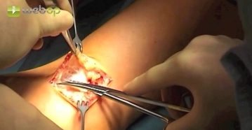
    Locate and dissect the cephalic vein, which is encircled centrally with a vessel loop. Close off the vein peripherally by simple ligation approximately 1 cm distal to the junction of the side branch. Occlude the vein centrad with a soft bulldog clamp and take down both the vein and its lateral branch peripherally. Carry the incision of the vein into the lateral branch, resulting in a flared shape.

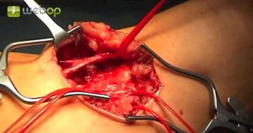
    After careful dilation probe the cephalic vein centrad with a Fogarty catheter to verify venous patency in the upper arm. Delicately dilate the vein when withdrawing the catheter. Administer 1000 IU of heparin saline solution centrad

    Tips:

    1. Do not bruise the vein with the forceps.

    2. If the peripheral vein is supplied by a suitable collateral branch, use it fashion a flared anastomosis. This will facilitate the  anastomosis.

    3. In case of small vein caliber, carefully dilate the cephalic vein along the entire course on the upper arm with a balloon catheter.

  3. Dissecting the brachial artery

    Video
     Dissecting the brachial artery

    Divide the aponeurosis of the biceps muscle of the arm longitudinally over the palpable brachial artery. Expose the artery while sparing its lateral branches, and encicrcle it first centrad and then peripherally with vessel loops.

    Tips:

    1. The median nerve parallels the brachial artery and is located below the center of the aponeurosis. Since at times the nerve runs rather superficially, the aponeurosis should be divided in small steps, perhaps even over a previously inserted Overholt forceps.

    2. Spare the small collateral branches of the brachial artery, possibly by clipping them temporarily.

Clamping the brachial artery and local TEA

After clamping the brachial artery, open up the vessel with a longitudinal incision. As the vessel

Activate now and continue learning straight away.

Single Access

Activation of this course for 3 days.

€4.99 inclusive VAT

Most popular offer

webop - Savings Flex

Combine our learning modules flexibly and save up to 50%.

from €3.29 / module

€39.50 / yearly payment

price overview

vascular surgery

Unlock all courses in this module.

€7.42 / month

€89.00 / yearly payment