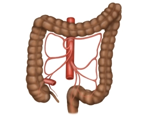
The colon is divided into the cecum with vermiform appendix, ascending colon, transverse colon, descending colon, sigmoid colon, and rectum.
The colon originates in the right iliac fossa and merges with the rectum at the level of the second sacral vertebra. The colon frames the loops of small bowel in inverted U-fashion.
Blood supply
Ascending and transverse colon are supplied by the ileocolic, right colic, and middle colic arteries, which originate on the right side of the superior mesenteric artery. To its right, the middle colic artery communicates with a branch of the right colic artery, and to its left with a branch of the left colic artery.
The descending and sigmoid colon and most of the rectum receive its blood supply from the inferior mesenteric artery via the left colic, sigmoid, and superior rectal arteries.
Anastomosis of Riolan
This anastomosis between the middle and left colic arteries, i.e., between the superior and inferior mesenteric arteries, is not always present. It can provide collateral arterial blood supply to the colon if one of these two mesenteric vessels is occluded.
Overview Arterial blood supply to the colon
Veins
The arteries are paralleled by the homonymous veins draining the blood into the superior and inferior mesenteric veins. Both vessels empty into the hepatic