Sigma resection, laparoscopic

  1. Creation of pneumoperitoneum / Placement of working trocars

    Video
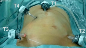
    Creation of pneumoperitoneum / Placement of working trocars

    The skin is incised in the midline approximately 3 cm cranial to the navel, the camera trocar (T1) is introduced into the abdominal cavity via a mini-laparotomy, and the pneumoperitoneum is established. After an orienting inspection of the abdominal cavity, the working trocars (T3, T4, T2) are introduced under direct vision.

    Note:

    The risk of injury to major intra-abdominal vessels by using a Veress needle to establish the pneumoperitoneum must be considered.

  2. Mobilization of the Sigmoid Colon

    Video

    The patient is placed in the right lateral position.
    The embryonic adhesions between the peritoneum and the sigmoid colon are dissected towards the left flexure on Gerota's fascia. The left ureter and the left iliac vessels must be clearly identified in this process.

  3. Mobilization of the left colonic flexure

    Video

    The patient is now in the anti-Trendelenburg and right lateral position. Initially, mobilization of the descending colon and left flexure from the lateral side is performed, for which the descending colon is stretched medially and the adhesions of the intestine to the Gerota's fascia are gradually dissected up to the level of the left flexure.

    To resolve the left flexure, the greater omentum is dissected starting from the middle of the transverse colon, providing access to the omental bursa. This involves the transection of the splenocolic ligament and the pancreatic-colonic connections. Subsequently, the left colon, including the flexure, is detached from all dorsal structures, allowing for a tension-free anastomosis.

    Tips:

    • Excessive pulling on the intestine can lead to lesions of the splenic capsule.
    • For the dissection, the Trendelenburg position ("head down") should be discontinued, while maintaining the right tilt of the operating table.
  4. Tubular dissection of the mesosigmoid

    Video
    Tubular dissection of the mesosigmoid

    The anti-Trendelenburg and right lateral positions are reversed. The mesosigmoid is incised medially, the retractor is introduced, and the mesentery of the sigmoid colon is transected close to the bowel with continuous exposure by ventral traction. The preparation should extend distally to the high-pull zone of the rectosigmoid junction (marked by the disappearance of the appendices epiploicae and the taeniae). Dissection along the colon ensures the intended preservation of the inferior mesenteric artery and its main branches. Caution: Identification of the ureter and the ovarian/testicular vessels.

Transection of the upper rectum

The resection planes are determined. These should lie orally beyond the inflamed or diverticulum-be

Activate now and continue learning straight away.

Single Access

Activation of this course for 3 days.

€7.99 inclusive VAT

Most popular offer

webop - Savings Flex

Combine our learning modules flexibly and save up to 50%.

from €3.70 / module

€44.50 / yearly payment

price overview

general and visceral surgery

Unlock all courses in this module.

€12.42 / month

€149.00 / yearly payment