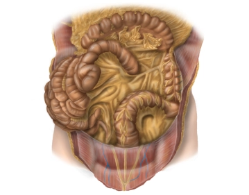
- Descending colon:
- Location: secondarily retroperitoneal
- Peritoneal suspension: fused with the posterior wall of the abdominal cavity
- Course: from the apex of the left flexure to the iliac fossa, connects to the transverse colon and transitions into the sigmoid colon.
- Length: 20-30 cm
- Sigmoid colon:
- Location: intraperitoneal
- Peritoneal suspension: sigmoid mesocolon
- Course: from the iliac fossa as a loop (S-shaped) to the level of the 2nd-3rd sacral vertebrae, connects to the descending colon and transitions into the rectum.
- Length: variable (elongated sigmoid), typically about 35 cm
-
Anatomy of the descending colon and sigmoid colon
![Anatomy of the descending colon and sigmoid colon]()
-
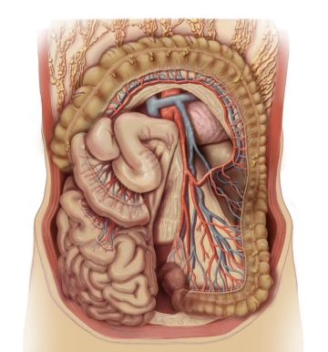
Vascular supply, lymphatic drainage, and nerves
![Vascular supply, lymphatic drainage, and nerves]()
- Arterial supply of the left hemicolon, sigmoid colon to the upper rectum by the inferior mesenteric artery
- Origin of the inferior mesenteric artery from the abdominal aorta at the level of the lumbar vertebra
Note: The inferior mesenteric plexus (inferior mesenteric ganglion) surrounds the origin of the inferior mesenteric artery
- Left colic artery: supplies the descending colon, an ascending branch anastomoses with the middle colic artery, a descending branch anastomoses with a sigmoid artery.
- Sigmoid arteries: 2-4 arteries, several small branches to the sigmoid colon, anastomoses to the left colic artery and superior rectal artery.
- Superior rectal artery: Travels dorsally to the upper rectum, anastomoses with the sigmoid artery and middle rectal artery from the internal iliac artery.
- Left colonic flexure: Watershed area between the supply territories of the superior mesenteric artery and the inferior mesenteric artery.
- Riolan's anastomosis: distant arcade between the superior mesenteric artery (middle colic artery) and inferior mesenteric artery (left colic artery) near the left colic flexure
Note: Inconstant! Riolan's anastomosis is not or not sufficiently developed in 20% of cases.
- Drummond's anastomosis: Vascular arcade that connects the colonic branches of the superior mesenteric artery and the inferior mesenteric artery close to the intestine. It forms a network mainly between the intestinal or marginal branches of the colonic arteries, which is why it is also called the marginal artery.
- Venous drainage via the left colic vein and sigmoid veins into the inferior mesenteric vein, which drains into the splenic vein behind the pancreatic tail. This forms the portal vein with the superior mesenteric vein and other visceral veins behind the pancreatic head.
- Lymphatic drainage along the inferior mesenteric artery (left colic lymph nodes, sigmoid lymph nodes)
- Important neural structures:
- Inferior mesenteric plexus (inferior mesenteric ganglion) (autonomic nervous system) at the origin of the inferior mesenteric artery
- Involved sympathetic nerves: lumbar splanchnic nerves, involved parasympathetic nerves: pelvic splanchnic nerves, both run to the inferior hypogastric plexus.
- Arterial supply of the left hemicolon, sigmoid colon to the upper rectum by the inferior mesenteric artery
Topographical Relationships
The descending colon runs from cranial to caudal adjacent to the left lateral abdominal wall. It ha
The descending colon runs from cranial to caudal adjacent to the left lateral abdominal wall. It ha
Activate now and continue learning straight away.
Single Access
Activation of this course for 3 days.
US$9.10
inclusive VAT
Most popular offer
webop - Savings Flex
Combine our learning modules flexibly and save up to 50%.
from US$7.04 / module
US$84.52/ yearly payment
general and visceral surgery
Unlock all courses in this module.
US$14.08
/ month
US$169.00 / yearly payment