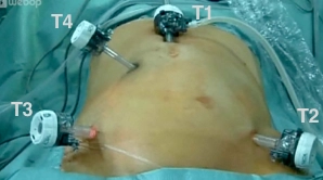
Sigmoidectomy, laparoscopic - general and visceral surgery
You have not purchased a license - paywall is active: to the product selection
Creating the pneumoperitoneum and placing the trocars
Make a medial skin incision about 3cm superior to the umbilicus. Introduce the trocar for the laparoscope (T1) into the abdominal cavity through a minilaparotomy and create the pneumoperitoneum. After gross inspection of the abdominal cavity, introduce the working trocars (T3, T3, T2) under direct view.
Note:
Be aware that creating the pneumoperitoneum with a Veress needle may risk injury to large intraabdominal blood vessels.
Mobilizing the sigmoid
Mobilizing the splenic flexure
Reposition the patient to reverse Trendelenburg while still in right lateral recumbent. Start freeing the descending colon and splenic flexure laterally from the left. In doing so, pull the descending colon mediad and successively divide the colonic adhesion with the Gerota fascia.
Starting in the middle of the transverse colon dissect the greater omentum off it, which will free the splenic flexure and open up the lesser sac. During this dissection the splenocolic ligament and attachments between the pancreas and colon will also be divided. Finally, the left colon including its splenic flexure has been freed from all posterior structures, allowing a tension-free anastomosis.
Tips:
- Abrupt traction on the colon may result in injury to the spleen.
- Dissection is facilitated by bringing the OR table from Trendelenburg (head low) back to level while still keeping the patient in right lateral recumbent.
Tubular Dissection of the mesosigmoid
Dividing the upper rectum
Extracorporeal sigmoid transection
Inserting the anvil and interiorizing the bowel
Colonic anastomosis
Single Access
Access to this lecture
for 3 days
€4.99 inclusive VAT

webop-Account Single
full access to all lectures
price per month
for the modul: vascular surgery