Start your free 3-day trial — no credit card required, full access included

Perioperative management - Umbilical hernia repair, open, preperitoneal umbilical mesh plasty (“PUMP”-Repair)

  1. Indications

    Considering an incarceration rate of 30 % with a lethality of up to 15 %, there is no doubt about the indication for surgical treatment of ventral abdominal wall hernias. An exception are small umbilical hernias whose fascial gap is less than 0.5 cm and therefore have only a low risk of incarceration.

    Hernia repair by direct suture is possible according to the guidelines of the European and American Hernia Society up to a hernia gap diameter of < 1 cm, provided there is no obesity (BMI > 30) and/or rectus diastasis.

    EHS (European Hernia Society) and AHS (American Hernia Society) see the limit for the use of a mesh at > 1 cm. Only for defects of 0 - < 1 cm should a suture procedure be chosen with a margin of discretion for defects between 1 - 2 cm. Up to 4 cm, a preperitoneal mesh plasty is recommended, alternatively minimally invasive sublay techniques (E/Milos, eTEP, TES) are also used, especially in the presence of rectus diastasis.

    Round self-expanding meshes are obsolete because no broad parietalization, which is only possible laparoscopically, occurs. The reason is the anatomy of the plicae converging in the umbilicus, which prevent the unfolding of these meshes. The preperitoneal space is the actual optimal layer for umbilical meshes.

    Presented in the article is the open preperitoneal, umbilical mesh plasty = PUMP-Repair.

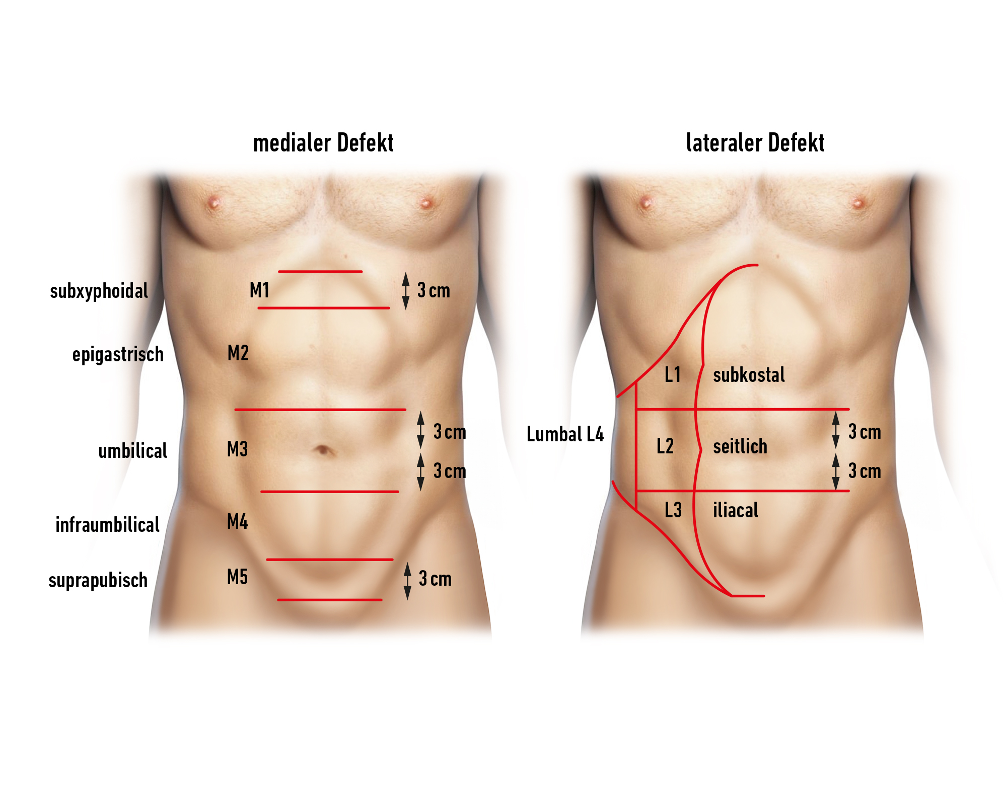
    This is a preperitoneal mesh technique with defect closure after insertion of a mesh that should overlap the defect by 3 cm on all sides. Therefore, the peritoneum must be dissected from the abdominal wall all around the defect by 3 cm on all sides.

    In the open repair of ventral hernias, the hernia orifice is basically only stretched, but not surgically enlarged. An enlargement of the hernia orifice would transform a primary hernia into an incisional one with a significantly higher recurrence rate.

    In obesity, data with lower wound complication rates are available for the lap IPOM compared to open procedures.

    For defects > 4 cm, proceed as with incisional hernias.

  2. Contraindications

    • For elective procedures, infection-free skin conditions are mandatory; pressure ulcers and superficial skin infections should initially be treated primarily conservatively.
    • The indication for hernia repair in patients with liver cirrhosis and ascites should be critically weighed; if necessary, preoperative optimization of liver function should be considered. In cases of severe coagulation disorders (Quick < 50 %, PTT > 60 s, platelets < 50 / nl) and pronounced portal hypertension with caput medusae, surgery should be avoided, especially due to the risk of uncontrollable bleeding from abdominal wall vessels.
    • A good respiratory situation, not compromised by acute infections, is also important. In case of respiratory infections, an elective procedure must be postponed.
  3. Preoperative Diagnostics

    History

    • Duration and progression of the hernia
    • In case of rapid increase in size, a pathological intra-abdominal event must be excluded!

    Clinical Examination

    The abdominal wall hernia is a clinical diagnosis and can usually be easily recognized in the standing patient. It is advisable to additionally examine the patient in a relaxed, lying position. If the patient is asked to lift the upper body, the fascial edge, the extent of the fascial defect, and the surrounding muscles can usually be assessed in reducible incisional hernias.

    Ultrasound of the Abdominal Wall

    • entire midline from xiphoid to lower abdomen
    • Condition of the linea alba: further fascial defects, rectus diastasis
    • CT only in special cases

     Classification of primary ventral hernias


    Small (S)

    Medium (M)

    Large (L)


    < 2 cm 

    ≥ 2 - < 4 cm

    ≥ 4 cm 

    Midline

    Epigastric


    Umbilical


    Lateral

    Spigelian


    Lumbar


     Classification of secondary ventral hernias (incisional hernias)

    Narbenhernie_PM.jpg

    The classification of secondary abdominal wall hernias is initially based on a medial or lateral defect location in the abdominal wall.

    The defect location of medial hernias is then more precisely delimited into subxiphoid, epigastric, umbilical, infraumbilical, and suprapubic. Laterally, the defects are divided into subcostal, lateral, iliac, and lumbar.

    Further consideration is given to the defect width of the incisional hernias: W1 (< 4 cm), W2 (4 - 10 cm) and W3 (> 10 cm).

    If multiple hernia defects exist (gridiron hernia, Swiss-cheese hernia), their size is determined by the total length and width.

  4. Special Preparation

    • Control of infection situations
    • Medication management in immunosuppression or anticoagulation
    • Control of cardiac and pulmonary risk factors
    • Single-shot antibiotic i.v. perioperatively (due to use of foreign material/mesh) possibly continuation of therapy in case of intraoperative signs of inflammation or bacterial contamination.
  5. Informed Consent

    • Wound healing disorders
    • Recurrence
    • Hematomas, secondary bleedings
    • Bowel injury
    • Injury to adjacent structures
    • Postoperative ileus
    • Infection
    • Thrombosis
    • Embolism
    • Reoperation
    • Temporary postoperative activity restriction
    • Chronic pain syndrome
    • Umbilical skin necrosis
  6. Anesthesia

  7. Positioning

    Positioning
    • Supine position, if necessary one arm abducted
  8. OR Setup

    OR Setup

    The surgeon stands to the right of the patient, the assistant stands opposite him.
    The instrumenting OR nurse stands at the foot end on the side of the assistant.

  9. Special Instrument Sets and Holding Systems

    Implant:

    • Non-resorbable, large-pored, surface-reduced plastic mesh
    • Alternatively, as in the film example, use of a bio-mesh
  10. Postoperative Treatment

    Postoperative Analgesia
    Non-steroidal anti-inflammatory drugs are generally sufficient; if necessary, escalation with opioid-containing analgesics can occur.
    Follow the links here to PROSPECT (Procedure Specific Postoperative Pain Management) and the current Guidelines for the Treatment of Acute Perioperative and Posttraumatic Pain.

    Medical Follow-up
    Inform patients about the reduced load-bearing capacity of the abdominal wall!

    Thromboembolism Prophylaxis
    In the absence of contraindications, due to the moderate thromboembolism risk (surgical procedure > 30 min duration), in addition to physical measures, low-molecular-weight heparin should be administered in prophylactic, possibly weight- or disposition risk-adapted dosage until full mobilization is achieved.
    To be noted: Renal function, HIT II (history, platelet control)
    Follow the link here to the current Guideline Prophylaxis of Venous Thromboembolism (VTE)

    Mobilization
    Immediate
    Resumption of normal physical activities after completion of wound healing.
    After 3 - 4 weeks, heavier physical exertion and sports should be possible again.

    Physiotherapy
    Not required

    Diet Build-up
    Immediate, without restriction

    Stool Regulation
    Not required

    Inability to Work
    Depending on the occupation 7 – 14 days