Are you sure you want to perform this action?
Peritoneal dialysis: Laparoscopic-assisted catheter insertion in CAPD
You have not purchased a license - paywall is active: to the product selection
Single Access
Access to this lecture
for 3 days
€4.99 inclusive VAT

webop-Account Single
full access to all lectures
price per month
for the modul: vascular surgery
from 8,17 €
-
Surgical anatomy of the anterior abdominal wall
Superficial layer
The superficial layer of the abdominal wall comprises the skin and the fatty tissue underneath (panniculus adiposus telae subcutaneae abdominis).
Middle layer
The middle layer primarily comprises the anterior and posterior abdominal muscles with their fascias.
Anterior abdominal muscles and rectus sheath
The anterior abdominal muscles comprise three rather flat muscles and the rectus abdominis. Anteromedially the flat muscles fuse with the rectus sheath and insert there with a broad tendon (aponeurosis). The muscles course in the following anteroposterior order:
- External oblique: Posteriorly, it originates at the thoracolumbar fascia and the inferior 7 ribs and then courses as anterior lamina of the rectus sheath to the median linea alba and the iliac crest of the pelvis. Its oblique fibers run superolaterally to inferomedially.
- Internal oblique: It courses from the linea alba to the iliac crest and the anterior margin of the pubic bone. Its oblique fibers run superomedially to inferolaterally (continuing the contralateral external oblique). Thus, both muscles crisscross obliquely in the anterior abdominal wall. Superior to the arcuate line, the internal oblique fuses with both the anterior and posterior lamina of the rectus sheath, and inferior to the arcuate line only with the anterior lamina.
- Transversus abdominis: Its fibers course anteriorly from the thoracolumbar fascia or the cartilage of the inferior ribs and the pelvis to the linea alba. In the superior region of the anterior abdominal wall it primarily constitutes the posterior lamina of the rectus sheath. Together with the oblique abdominal muscles it constitutes the anterior lamina inferior to the arcuate line. The transversalis fascia constitutes the posterior wall of these three muscles.
- On both sides the rectus abdominis originates at the cartilage of ribs 5-7 and inserts in the pubic bone near the symphysis pubis. Tendinous intersections divide the long muscles into several bellies (“six-pack”). The pyramidalis muscle is an inconsistent muscle coursing anterior to the rectus abdominis and braces the linea alba. Thus, the rectus sheath is a tendinous canal investing the flat abdominal muscles and comprising the rectus abdominis and pyramidalis muscle as well as various vessels and nerves (inferior and superior epigastric artery and vein, intercostal nerves 5-12).
Function
For flexion and rotation of the trunk and abdominal straining both oblique abdominal muscles (m. obliquus externus and internus abdominis - oblique cross) and the rectus abdominis plus transversus abdominis (upright cross) brace the anterior abdominal wall in the fashion just described.
The cremaster muscle derives from the internal oblique and transversus abdominis. It is the muscular investment of the spermatic cord and can lift the testicles (cremasteric reflex).
Deep layer
The transversalis fascia is the deep posterior layer of the abdominal wall. As the most internal layer of connective tissue (only separated from the free abdominal cavity by the peritoneum), it covers the internal aspect of the rectus abdominis and transversus abdominis and conjoins with the arcuate line and inguinal ligament. The deep inguinal ring with the entry to the inguinal canal is situated inferolaterally.
Posterior muscles
The major posterior muscle of the abdominal wall is the quadratus lumborum, which courses below the transversus abdominis from the lowermost rib and costal processes of the lumbar spine to the iliac crest.
Blood supply and innervation
The arterial blood supply follows the above layers of the abdominal wall:
- The superficial and middle layers are supplied by the
- → inferior posterior intercostal arteries (including the subcostal artery),
- → superficial epigastric artery,
- → superficial circumflex iliac artery and the
- → external pudendal artery.
- The deep layer is supplied by the
- → lumbar arteries,
- → inferior epigastric artery,
- → deep circumflex iliac artery and the
- → iliolumbar artery.
The venous blood of the abdominal wall drains via veins (primarily → inferior vena cava) eponymic with their corresponding arteries:
Via the superficial epigastric vein (→ great saphenous vein) and the inferior epigastric vein (→ external iliac vein). The venous blood only enters the superior vena cava via the thoracoepigastric veins and the azygos and hemiazygos veins.
The abdominal wall is innervated by intercostal nerves and branches of the lumbar plexus:
- As noted above, the inferior intercostals (including the subcostal nerve) innervate the external oblique abdominal muscle and the rectus abdominis.
- The iliohypogastric nerve, originating at the lumbar plexus, innervates all anterior abdominal muscles, as does the ilioinguinal nerve except for the rectus abdominis, while the genitofemoral nerve supplies the transversus abdominis.
- The iliohypogastric and ilioinguinal nerves also course between the muscles innervated by them and supply the skin of the anterior abdominal wall.
Superior to the umbilicus, lymph from the anterior abdominal wall drains cephalad (into the axillary and parasternal lymph nodes), while inferior to the umbilicus it drains caudad (into the inguinal and iliac lymph nodes). Lymph from the lateral abdominal wall drains into the lumbar lymph nodes.
-
Peritoneal dialysis: Effects on the peritoneum and the resultant changes in morphology, function and clinical picture
Following are the possible modalities in renal replacement therapy:
- Peritoneal dialysis (PD)
- Hemodialysis (HD)
- Renal transplantation
While PD is reserved for acute dialysis in infants and toddlers and those situations where hemodialysis is not possible, it excels in chronic dialysis settings and today is the most common modality in home dialysis.
In CAPD (Continuous Ambulatory Peritoneal Dialysis) the patient changes the dialysate 3 – 5 times daily by draining spent dialysate from the abdominal cavity into an empty bag and instilling new dialysate. This takes about 20 minutes. The times when the dialysate must be changed can be adapted to the daily routine of the patient; usually, 3 – 4 changes are easily integrated into the daily routine and these times may vary by 1 – 2 hours.
Single Access
Access to this lecture
for 3 days
€4.99 inclusive VAT

webop-Account Single
full access to all lectures
price per month
for the modul: vascular surgery
from 8,17 €
Single Access
Access to this lecture
for 3 days
€4.99 inclusive VAT

webop-Account Single
full access to all lectures
price per month
for the modul: vascular surgery
from 8,17 €
-
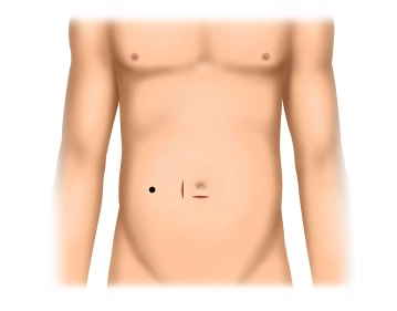
Infraumbilical access
-
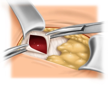
Exposure of the umbilical orifice
Whenever present, use the umbilical orifice for trocar insertion (as demonstrated in this video clip). To do so, circumvent the umbilicus with a finger or blunt instrument and expose its insertion at the level of the fascia. Here, the umbilicus is sharply dissected off its insertion, thereby exposing the umbilical orifice. Now clamp the edges of the fascia with Mikulicz clamps.
Tip:
In planned peritoneal dialysis, the umbilical orifice must be carefully closed because otherwise the dialysate instilled into the abdominal cavity may result in herniation. If there is no umbilical orifice which may double as trocar access, the latter is established as usual in laparoscopy.
-
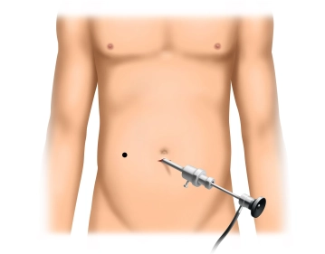
Initiation of pneumoperitoneum and inspection of the abdominal cavity
Insert the trocar for the camera through a small incision of the peritoneum and establish the pressure controlled pneumoperitoneum. Insert the camera and inspect the abdominal cavity, paying particular attention to any adhesions which may affect the choice on which side the catheter will be inserted. While the video clip demonstrates adhesions after previous open appendectomy, these do not have to be taken down and the catheter may be inserted on the right side.
-
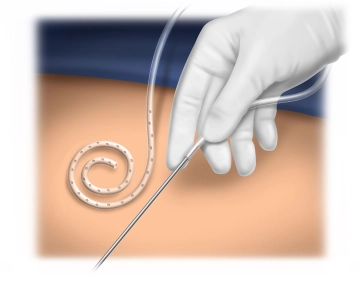
Threading the catheter
Slip the catheter over the stylet. Accidental twisting of the catheter during this maneuver is best prevented by keeping the visible marker line on the catheter straight.
Tips:
- It is easier to slip the catheter over the stylet once it has been flushed with or soaked in saline.
- The catheter coiling should always point laterally (i.e., in the video clip with insertion in the right lower quadrant to the right). If after insertion the catheter coiling is located medially, the catheter tends to reposition the coil laterally which may result in catheter dislocation from the lesser pelvis.
-
Pararectal access at the level of the umbilicus
Between the umbilicus and the planned catheter exit site incise the skin above the rectus muscle in the right lower quadrant. Transect the subcutis, expose and then split the anterior rectus sheath. Bluntly split the rectus muscle, and with a long-term absorbable suture preplace a purse-string suture on the posterior rectus sheath/peritoneum.
Tip:
A well-established pneumoperitoneum will prevent accidental capture of the greater omentum or intestines when placing the purse-string suture.
-
Catheter insertion
Insert the stylet with its mounted catheter through the posterior rectus sheath / peritoneum incised within the purse-string suture and place it in the lesser pelvis under laparoscopic view. Injuries in the lesser pelvis are best prevented by advancing the catheter with one hand while keeping the stylet stationary with the other hand. Remove the stylet once the catheter has been placed correctly. When tightening the purse-string suture ensure that the catheter cuff is in close contact with the peritoneum. Mount the free end of the catheter on a tunneling stylet (16 French) and advance the latter deep in the subcutis in curved fashion until it punches through the marked site of the planned catheter exit. Remove the stylet, mount the titanium adapter and attach the transfer unit. As a final step, inspect the peritoneum and catheter placement laparoscopically.
Tips:
- Before tightening the purse-string suture reduce the intraabdominal pressure of the pneumoperitoneum somewhat because this will prevent the rectus sheath and peritoneum from tearing while the suture is tied.
- If after tying the purse-string suture the catheter can still be pushed/pulled, but only with difficulty, it is well anchored and will not have become compressed.
- Once the tunneling stylet has emerged at the planned catheter exit site, check laparoscopically for possible iatrogenic perforation of the peritoneum by the stylet.
-
Infraumbilical suture of the peritoneum/fascia and catheter trial run
After a running infraumbilical suture of the peritoneum/fascia and definite closure of the umbilical orifice, test the patency of the catheter by instilling and then draining about 1.5 l dialysate (spiked with 5000 IU heparin). This maneuver also serves to detect any leakage at the peritoneal incisions.
-
Layered wound closure and refixation of the umbilicus
The pararectal incision of the right anterior rectus sheath is closed with a running suture, taking care not to compress the catheter. After resuturing the umbilicus to the fascia, close both access sites in layered fashion. Close the transfer unit with a cap that has been wetted with iodine disinfectant.
Single Access
Access to this lecture
for 3 days
€4.99 inclusive VAT

webop-Account Single
full access to all lectures
price per month
for the modul: vascular surgery
from 8,17 €
Single Access
Access to this lecture
for 3 days
€4.99 inclusive VAT

webop-Account Single
full access to all lectures
price per month
for the modul: vascular surgery
from 8,17 €
-
Literature summary
-
Ongoing trials on this topic
-
Reviews
-
References on this topic
-
literature search
Literature search under: http://www.pubmed.com