
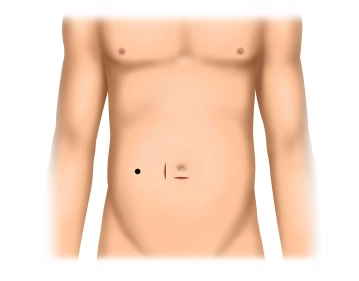
Infraumbilical skin incision, division of the subcutis and exposure of the fascia.
-
Infraumbilical Access
![Infraumbilical Access]()
Soundsettings -
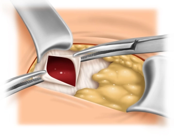
Exposure of the Umbilical Defect
![Exposure of the Umbilical Defect]()
Soundsettings If an umbilical defect is present (as in the video example), it is used as the trocar access. For this, blunt or digital circumscription of the umbilicus and exposure of its insertion at the fascial level is performed. Here, the umbilicus is sharply dissected and the umbilical defect is thus exposed. Subsequently, grasping of the fascial edges with Mikulicz clamps.
Tip:
Umbilical defects must be carefully repaired in planned peritoneal dialysis, as otherwise, due to the intra-abdominally instilled dialysate, hernia formation can occur. If no umbilical defect is present that can be used as trocar access, it is created as in any other laparoscopy.
Establishing the Pneumoperitoneum and Inspection of the Abdominal Cavity
Insertion of the optical trocar through a small incision of the peritoneum and pressure-controlled
Insertion of the optical trocar through a small incision of the peritoneum and pressure-controlled
Activate now and continue learning straight away.
Single Access
Activation of this course for 3 days.
US$9.10
inclusive VAT
Most popular offer
webop - Savings Flex
Combine our learning modules flexibly and save up to 50%.
from US$7.08 / module
US$85.05/ yearly payment
general and visceral surgery
Unlock all courses in this module.
US$14.17
/ month
US$170.10 / yearly payment
Webop is committed to education. That's why we offer all our content at a fair student rate.