Start your free 3-day trial — no credit card required, full access included

Perioperative management - Abdominal wall closure – techniques: Loop suture and small tissue bites

  1. Special anatomical and functional aspects of the anterior abdominal wall

    Special anatomical and functional aspects of the anterior abdominal wall

    The highly segmental structure of the chest wall structure continues to a lesser degree in the abdominal wall but decreases in the craniocaudad direction.

    Since the segmental character of the anterior part of the ventral abdominal wall is especially pronounced in the superior region, the forces there act primarily in the mediolateral direction. Midline incisions result in greater centrifugal tension at the fascial edges than do transverse incisions. Since the longitudinal strain becomes more prominent caudad to the umbilicus, there is less lateral tension on the fascial edges in longitudinal incisions; as a result, the abdominal wall craniad to the umbilicus has a greater tendency to wound dehiscence than does the inferior region.
    Closure of midline laparotomy is associated with high tensile stress, which could help explain the high rate of postoperative incisional hernias.

  2. Aspects of surgical wound closure in laparotomy

    The reconstructed abdominal wall with a mechanically stable laparotomy wound healed with satisfactory cosmesis is the visible expression of a successful operation.

    Good fascial healing is achieved not by abundant but rather by little and adequately matured scar tissue. This meets the surgical requirements for a gentle technique as well as avoidance of wound infections, and high suture tension.

    Incisional hernias are the most common long-term complication after laparotomy.

    Optimized fascial healing is the key for preventing incisional hernias.

    This requires the understanding that fascial suture only serves as a temporary support during local wound healing processes.
    During the exudative phase (days 1-4) the wound has no tensile strength. The proliferative phase (days 5-20) sees the development of granulation tissue and the emergence of a new connective tissue matrix, which, however, only has 15-30% of the original tensile strength. Hematoma or wound infection may significantly prolong this process.

    The required formation of scar tissue should not be considered as a static process, but rather as a chronic remodeling of the abdominal wall, even after years.

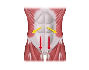
    Continuous mass closure of the abdominal wall with a suture to wound length ratio of at least 4:1 with monofilament, delayed-absorbable sutures is superior to the interrupted suture technique. Its benefit is better biomechanical properties with favorable collagen synthesis along the incision as well as the economic aspect of significant savings in time and material. One important biomechanical factor appears to be the distribution of suture tension across small tissue bridges.

    To prevent "buttonholes" from triggering incisional hernias, adequate tension of the running suture, adapting the edges of the fascia> with low strain on the tissue bridges, must be ensured during closure.

    In addition, the elasticity of the suture material, corresponding to the physiological excursions of the abdominal wall, is another factor in preventing "buttonholing".

  3. Suture material

    During healing, the abdominal wall is subjected to a wide variety of stressors. The sutures must absorb some of the resulting forces. Ideally, these sutures should have elastic properties resembling those of rubber. The repair phase of the abdominal wall requires about three months to achieve 75% of the original tissue strength. The loss of suture tensile strength should match the healing of the abdominal wall.

    The ideal suture material is a delayed-absorbable suture.

    Although non-absorbable sutures reduce the incidence of incisional hernia, they are associated with significantly more local complications such as suture fistulas and prolonged wound pain.

    MonoMax®, the suture employed in this presentation, belongs to a new generation suture material made of poly-4-hydroxybutyrate. MonoMax® is a monofilament high tensile strength suture which physically buttresses the abdominal wall for six months and is completely absorbed within one year. It excels with ultra-late absorption, high elasticity, and a high degree of flexibility. It is the elasticity which reduces the tendency of the stitch to cut through the tissue toward the edges of the fascia and thus protects the tissue.

    The looped sutures currently favored benefit from the fact that one of the knots in the corners of the wound may be dispensed with.

  4. “Small bites” technique

    At present, the technique of choice in secure fascial closure is a running looped suture 1 cm from the wound edges so as not to impair the active healing zone of 5 mm along the edges of the fascia.

    However, Israelsson et al. were able to demonstrate that compared to the "long stitch" group an innovative "short stitch" technique with a reduction in stitch distance to the wound edge from 10 mm to 5‑8 mm was able to reduce infection rates and the development of incisional hernia.

    This result is due to a further reduction of the tissue-damaging tensile forces in each stitch and to a large amount of "spare material" in the coils of the suture, preventing an inappropriate increase in suture tension when the abdomen is distended.

    This suture technique is characterized by the following:

    • Delayed-absorbable monofilament suture material size 0 or 2/0
    • Small needle HR 26 or HR 30
    • Running suture technique
    • Encompassing only the aponeurosis
    • Suture/incision length ratio of at least 5:1
    • Stitch interval 4‑5 mm
    • Distance from wound edge 5‑8 mm
    • Tension adapted to the suture line
    • Suture bridges should be visible
  5. Subcutaneous wound closure and skin sutures

    Subcutaneous sutures have long been a standard step in layered wound closure and was intended to prevent the formation of wound cavities where wound secretion can accumulate and become infected; they also help to prevent superficial wound dehiscence.

    At present, there is no general agreement on the importance of subcutaneous sutures and subcutaneous wound drains. The fact is that without suturing the subcutaneous layer before apposing the skin, this will leave a wound cavity of varying size which of course will fill with exudate. The thickness of the subcutaneous layer is also directly associated with the formation of wound infections. From a physiological perspective, the problems of a subcutaneous cavity must be weighed against the problem of introducing additional foreign material. Several trials have demonstrated a lower rate of subcutaneous hematomas, seromas, and wound infections after suturing the subcutaneous tissue.

    Although no evidence-based statements regarding the benefits of subcutaneous sutures and subcutaneous drains can be offered at this time, one or both measures appear to be reasonable in terms of pathophysiology.

    During skin closure, skin staples, of all the materials, appear to have the lowest infection rates in contaminated wounds and are also easy to apply.

    Although various trials have demonstrated that patient satisfaction with stapled skin sutures is worse than with skin sutures (especially with subcuticular sutures), objective measurements of long-term wound healing achieve the same results with both techniques.

  6. Abdominal wound dehiscence

    Abdominal wound dehiscence ("burst abdomen") is an early postoperative failure of the retaining function of the sutures and thus of the laparotomy closure.

    • Incidence: 0.5‑3 %
    • High mortality due to the severity of the underlying disease and pulmonary complications 
    • Confirmed etiological factors in multifactorial events include the pathological increase in intraabdominal pressure because of intestinal paralysis, ileus, peritonitis, and hematoma, as well as hypoalbuminemia
    • The most common pathomechanism involves the sutures cutting through the edge of the fascia into the incision.
    • It is treated by definitive closure or fashioning of a temporary pressure-relieving intestinal stoma.
    • Mass closure of the abdominal wall is superior to layered closure.