1. Anterior Abdominal Muscles
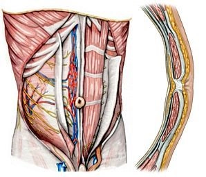
Rectus abdominis muscle: Straight abdominal muscle within the rectus sheath with 3-4 tendinous intersections (intersectiones tendineae) that are fused with the anterior layer of the rectus sheath.
Pyramidalis muscle: Originates from the superior pubic ramus and inserts into the linea alba, located ventrally to the rectus abdominis muscle in its own sheath within the anterior layer of the rectus sheath.
2. Layered Structure of the Anterior Abdominal Wall
Rectus sheath: Encloses the rectus abdominis muscle; above the midpoint between the navel and symphysis, it is differentiated into an anterior and a posterior layer; the posterior layer ends here in the form of the arcuate line; above this line, the external oblique muscle inserts into the anterior layer of the rectus sheath, the internal oblique muscle into both the anterior and posterior layers, and the transversus abdominis muscle into the posterior layer.
Linea semilunaris: Transition zone between the aponeuroses of the lateral abdominal muscles and the lateral edge of the rectus sheath.
Linea alba: Approximately 1 cm wide, firm connective tissue strip between the right and left rectus sheath, extending from the sternum to the symphysis.
Transversalis fascia: Above the arcuate line, it covers the posterior layer of the rectus sheath internally, below the line it lies directly on the rectus abdominis muscle.
3. Internal Relief of the Abdominal Wall
Median umbilical fold: Median peritoneal fold running from the navel to the bladder, containing the median umbilical ligament (fibrous cord = urachus remnant).
Medial umbilical fold: Paired peritoneal fold, containing on each side the medial umbilical ligament = obliterated remnant of the bilateral umbilical artery, umbilical artery.
Lateral umbilical fold: Paired peritoneal fold, beneath which lies on both sides the inferior epigastric artery with two accompanying veins each.
4. Conduits
a) Arteries
Superior epigastric artery: Continuation of the internal thoracic artery, anastomoses at the level of the navel with the inferior epigastric artery.
Inferior epigastric artery: Arises from the external iliac artery and runs like the aforementioned artery on the dorsal surface of the rectus abdominis muscle within the rectus sheath.
Superficial epigastric artery: Originates from the femoral artery and distributes after crossing the inguinal ligament in the subcutaneous tissue of the anterior abdominal wall.
Posterior intercostal arteries VI – XI and subcostal artery: Originate from the thoracic aorta; their terminal branches run obliquely downward between the internal oblique muscle and the transversus abdominis muscle and extend from the lateral side into the rectus sheath, where they anastomose with the superior and inferior epigastric arteries.
b) Veins
Superior epigastric veins: Accompany the artery of the same name; anastomose with branches of the inferior epigastric vein and drain into the internal thoracic veins.
Inferior epigastric vein: Branches into accompanying veins of the inferior epigastric artery and drains into the external iliac vein.
Superficial epigastric vein: Runs parallel to the artery of the same name (see above).
c) Lymphatic Vessels
Superficial lymphatic vessels: Above the navel, they lead to the axillary lymph nodes, below to the inguinal lymph nodes.
Deep lymphatic vessels: Generally run parallel with the blood vessels; reach the parasternal, lumbar, and external iliac lymph nodes.
d) Nerves
Intercostal nerves VI – XII: As ventral branches of the thoracic nerves VI – XII; they enter the abdominal wall behind the costal cartilages between the internal oblique muscle and the transversus abdominis muscle; motor branches supply the anterior and lateral abdominal muscles, the sensory branches the abdominal skin.
Iliohypogastric nerve, ilioinguinal nerve, and genitofemoral nerve: Participate in the motor and sensory innervation of the lower abdominal region and genitalia.