Start your free 3-day trial — no credit card required, full access included

Left hemicolectomy, ArtiSential-assisted

  1. Positioning

    Positioning

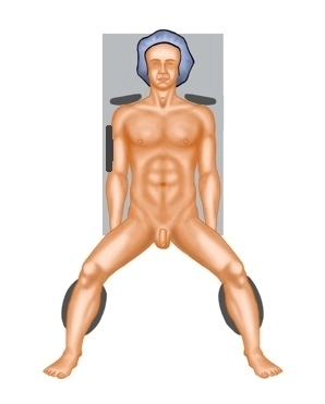
    Ideally, positioning is done in lithotomy position on a large vacuum cushion (on the right side, the cushion supports the rib cage and iliac crest to prevent the patient's weight in right lateral position from pressing on the arm).

    Additionally, sufficient support of the shoulders must be ensured to prevent slipping during prolonged Trendelenburg and right lateral positioning.

    It is recommended to position both arms (caution: cotton wrapping when positioning with a cloth sling).

    Cotton wrapping of the knees and proximal lower legs is also performed to prevent pressure injuries.

    For leg positioning, padded "boots" are recommended for optimal cushioning with separate sterile draping of the legs. Alternatively, the legs can be positioned in leg holders with fixation in these.

    The legs should be adjustable in flexion and extension via the OR table control.

    Note: Before sterile washing, always test the positioning in Trendelenburg and right lateral position with the remote control.

    Caution: Vacuum cushions may have leaks. Therefore, they should be checked again before sterile draping.

  2. Creation of Capnoperitoneum and Trocar Positioning

    Creation of Capnoperitoneum and Trocar Positioning

    The skin is incised in the midline through an infraumbilical incision, and the camera trocar is introduced into the abdominal cavity via a minilaparotomy to establish the capnoperitoneum. After an orienting inspection of the abdominal cavity, the working trocars are introduced under direct vision. An 8mm trocar is placed in the midclavicular line supraumbilically, a 12mm trocar in the midclavicular line in the right lower abdomen, a 5mm trocar for the assistant in the epigastrium, and another 12mm trocar in the left lower abdomen.

    Note: The creation of the capnoperitoneum varies among different clinics. Alternatively, this can be done using a Veress needle or a First Entry trocar instead of the open approach. In the latter two methods, utmost caution must always be exercised to avoid the risk of injuring major intra-abdominal vessels.

  3. Preparation and Transection of the Inferior Mesenteric Vein

    Video
    Preparation and Transection of the Inferior Mesenteric Vein
    Soundsettings

    After shifting the small intestine to the right and exposing the ligament of Treitz, preparation is performed in the right lateral position and Trendelenburg position using the Artisential bipolar forceps and the articulating spatula (Livsmed Artisential Monopolar spatula). The spatula is used to incise the serosa medially and below the inferior mesenteric vein (so-called Infra IMV-Approach). This allows entry into the avascular plane between the mesocolon and the retroperitoneum in front of the Gerota's fascia.

    Note: The proximal jejunum is tensioned with an atraumatic grasping forceps through the assistant trocar.

    The skeletonization of the inferior mesenteric vein at the ligament of Treitz is performed immediately at the lower edge of the pancreas. The vein is closed with 3 absorbable locking vascular clips, with 2 placed proximally, and then transected between the clips.

    Note: An explanation of the articulated instruments used from Livsmed Artisential can be found in the section "Perioperative Management".

  4. Mobilization medial to lateral

    Mobilization medial to lateral
    Soundsettings

    Detachment of the last adhesions to the duodenojejunal flexure. Further dissection in the avascular plane between the colon mesentery and retroperitoneum in front of the Gerota's fascia, primarily by blunt dissection of the Gerota's fascia and occasional use of monopolar current. Proceeding further caudally, progressively detaching the colon mesentery and mobilizing from medial to lateral. The inferior mesenteric vein serves as a guiding structure.

    Note. It is important to pull the colon mesentery ventrally. Through the assistant trocar, the mesentery is tented during the medial to lateral dissection.

  5. Preparation and Transection of the Inferior Mesenteric Artery

    Preparation and Transection of the Inferior Mesenteric Artery
    Soundsettings

    Trendelenburg position. Incision of the serosa below the inferior mesenteric artery from the medial side (Infra IMA Approach). The correct site for the incision is identified by a line between whitish fat (retroperitoneum) and yellowish fat (mesocolon). Entry into the spider web layer in Waldeyer's space. Connection of the two incision lines. Exposure and skeletonization of the inferior mesenteric artery. The artery is secured with 3 absorbable locking clips and transected between them. In this process, 2 clips remain in situ and one is located on the specimen.

     Note: The transection is performed proximal, one centimeter after the artery's origin from the aorta, to preserve the nerves (inferior mesenteric plexus).

  6. Mobilization medial to lateral

    Mobilization medial to lateral
    Soundsettings

    Further dissection from medial to lateral under elevation of the mesocolon. Extensive exposure and careful preservation of the ureter. Finally, the tumor in the descending colon is also exposed coming from the medial side. The dorsal dissection is performed with a sufficient safety margin retroperitoneally.

Preparation of the Rectosigmoid Junction

Tension of the rectosigmoid junction ventrally and entry into Waldeyer's space. The correct layer i

Activate now and continue learning straight away.

Single Access

Activation of this course for 3 days.

US$9.30  inclusive VAT

Most popular offer

webop - Savings Flex

Combine our learning modules flexibly and save up to 50%.

from US$7.19 / module

US$86.33/ yearly payment

price overview

general and visceral surgery

Unlock all courses in this module.

US$14.38 / month

US$172.70 / yearly payment