
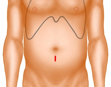
After sterile washing and draping of the operative field, the mini-laparotomy is performed immediately below the umbilicus. Creation of the pneumoperitoneum via the Veress needle.
-
Minilaparotomy
![Minilaparotomy]()
Soundsettings -
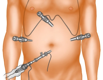
Positioning of the Trocars
![Positioning of the Trocars]()
Soundsettings Insertion of an 11-mm optical trocar; Placement of the additional 11-mm working trocars left lateral (working trocar e.g. for the scissors) and in the epigastrium (working trocar e.g. for a grasping forceps). A 5-mm trocar is placed in the right upper quadrant (working trocar for e.g. the suction device).
Exploration
Visualization of the cyst, opening of the cyst with scissors through electrocoagulation. Aspiration
Visualization of the cyst, opening of the cyst with scissors through electrocoagulation. Aspiration
Activate now and continue learning straight away.
Single Access
Activation of this course for 3 days.
US$9.30
inclusive VAT
Most popular offer
webop - Savings Flex
Combine our learning modules flexibly and save up to 50%.
from US$7.23 / module
US$86.85/ yearly payment
general and visceral surgery
Unlock all courses in this module.
US$14.47
/ month
US$173.70 / yearly payment
Webop is committed to education. That's why we offer all our content at a fair student rate.