Start your free 3-day trial — no credit card required, full access included

Incisional hernia repair, open sublay

  1. Findings

    Video
    Findings
    Soundsettings

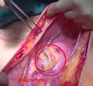
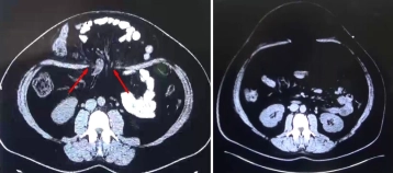
    In the young patient, a massive abdominal wall hernia had developed postpartum – without prior abdominal intervention – which led to the displacement of large intestinal segments into the hernia sac (left CT image with the marked hernia orifice).
    In advanced eventration of the viscera through the fascial defect, it was decided preoperatively to condition the abdominal wall with a progressive pneumoperitoneum. This measure resulted in a complete spontaneous repositioning of the hernia sac contents.

    In case of incomplete family planning for the patient, mesh augmentation with a biomesh in sublay technique is indicated, as this mesh exhibits better extensibility and is remodeled into autologous tissue.

  2. Skin Incision and Opening of the Hernia Sac

    Skin Incision and Opening of the Hernia Sac
    Soundsettings

    Access is achieved via a median upper abdominal incision extending below the navel. Opening of the hernia sac right at the beginning to identify adhesions and facilitate entry into the abdominal cavity. In the depth, the hernia orifice is visible, which is a large midline defect in the upper abdomen.

  3. Exposure of the Fascial Defect

    Soundsettings

    Exposure of the fascia at the hernia orifice by dissecting the subcutis. Incision of the hernia sac and initial preservation of the tissue, to possibly use it in the reconstruction phase. The thinned linea alba is opened cranially and caudally until sufficiently stable tissue is reached. The exploration of the abdomen remains unremarkable, there are no adhesions.

    Note: In the absence of a history of passage problems/recurrent ileus conditions, interenteric adhesiolysis can generally be omitted.

  4. Epifascial Preparation; Hernial Sac Resection

    Soundsettings

    Sparing epifascial preparation with resection of the hernial sac from the subcutis.

Incision of the Rectus Sheath

By pulling on the fascial edge  the posterior rectus sheath is visualized and opened close to the e

Activate now and continue learning straight away.

Most popular offer

webop - Savings Flex

Combine our learning modules flexibly and save up to 50%.

from US$7.19 / module

US$86.33/ yearly payment

price overview

general and visceral surgery

Unlock all courses in this module.

US$14.38 / month

US$172.70 / yearly payment