Start your free 3-day trial — no credit card required, full access included

Perioperative management - Incisional hernia surgery, laparoscopic with Ventralight™ ST mesh and ECHO PS™ positioning system

  1. Indications

    According to the guidelines of the EHS and AHS, the laparoscopic IPOM technique is recommended for larger primary and secondary abdominal wall hernias and in patients with an increased risk of wound complications. This particularly concerns patients with obesity (BMI >_ 30) and patients with a defect size of over 4 cm. However, the defect should not exceed a size of 8 - 10 cm.

    The laparoscopic intraperitoneal mesh implantation currently represents the most common endoscopic, minimally invasive technique for the treatment of abdominal hernias worldwide. In Germany, however, there is a decline in this method due to fear of adhesiogenesis with the formation of prosthetic-intestinal fistulas despite improved meshes regarding their coating. Other reasons include an increased rate of acute and chronic postoperative pain, presumably because the method is expensive. Nevertheless, it is an important alternative technique.

    Secondary abdominal wall hernias:

    Incisional hernia is the most common complication after laparotomy with a prevalence between 3 and 40% regardless of the abdominal wall closure chosen.

    Risk factors for the development of incisional hernias:
    BMI >­_ 25, male gender, recurrent incision, malignant disease, wound contamination, open surgery, COPD, positive family history.

    When determining the indication, the previous intervention should be at least 6 months ago.

    Closure of the hernia defect:

    There are studies that recommend an additional closure of the hernia defect in IPOM to reduce the rate of recurrences, seroma formation, and pseudo-recurrences. In the case of a small defect (< 3cm), a direct closure of the defect is used, and for larger defects, a so-called LIRA (intracorporeal rectus aponeuroplasty) with a knotless suture is applied. In a recent study (Pizza F et al 2023), the superiority of a hernia defect closure could not be confirmed.

  2. Contraindications

    • Relative contraindication for large defects (>10 cm) due to increased pain and recurrence rate
    • Giant hernia with no access for trocars (outside the planned mesh position!)
    • Split-thickness skin graft-covered former laparostoma/necrosis
    • Bacterial peritonitis (foreign material/mesh not in inflammation)
    • Ileus due to the risk of bowel perforation
    • Decompensated cardiorespiratory insufficiency
    • In severe cardiac and pulmonary pre-existing conditions, the benefits and risks of laparoscopy must be weighed, but even in higher-grade heart failure or respiratory impairment, laparoscopy under monitoring (blood pressure, pulse, ECG, oxygen saturation) is feasible.
    • Severe coagulation disorders (Quick < 50%, PTT > 60 s, platelets < 50 /nl) and pronounced portal hypertension with caput medusae, in both cases mainly due to the risk of bleeding from abdominal wall vessels.
  3. Preoperative Diagnostics

    • The abdominal wall hernia is a clinical diagnosis and can usually be well recognized in a standing patient. It is advisable to also examine the patient in a relaxed, lying position. When the patient is asked to raise the upper body, the fascial edge, the extent of the fascial defect, and the surrounding muscles can usually be assessed in reducible incisional hernias.
    • For smaller primary hernias, abdominal ultrasonography is a meaningful imaging procedure.
    • To determine the defect location and extent, especially in incisional hernias, and to depict the abdominal wall anatomy, CT is the best diagnostic procedure; alternatively, an MRI can be performed.
    • In cases of previous incisional hernia repairs, a corresponding operative report is often helpful, especially if a mesh repair has already been performed. Here, in addition to the exact surgical technique (extra- or intraperitoneal mesh placement, augmentation, or bridging of the fascial defect), the type of mesh material is particularly important.
    • In extensive findings, thorough cardiopulmonary functional diagnostics are necessary due to the increase in pressure after repositioning the eventrated viscera.

    EHS (European Hernia Society) Classification

    To better characterize the present hernia, the classification of the EHS should be used. 

    Classification of Primary Ventral Hernias


    Small (S)

    Medium (M)

    Large (L)


    < 2 cm 

    ≥ 2 - < 4 cm

    ≥ 4 cm 

    Midline

    Epigastric


    Umbilical


    Lateral

    Spigelian


    Lumbar


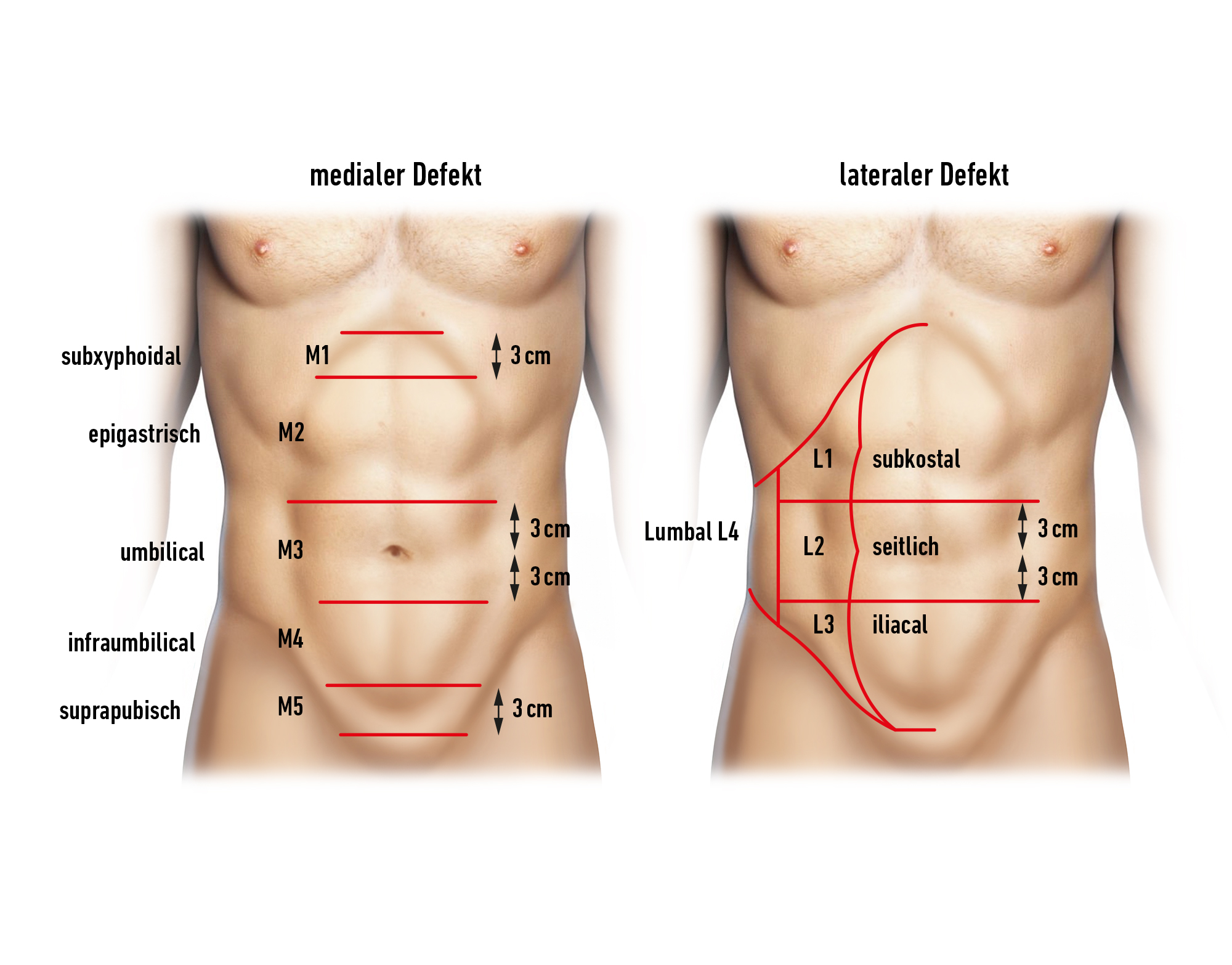
    The Classification of Secondary Abdominal Wall Hernias is initially based on a medial or lateral defect location in the abdominal wall.

    70-PM-3

    The defect location of medial hernias is then more precisely defined as subxiphoidal, epigastric, umbilical, infraumbilical, and suprapubic. Laterally, the defects are classified as subcostal, lateral, iliac, and lumbar.

    Further consideration is given to the defect width of incisional hernias: W1 (<4cm), W2 (4-10cm), and W3 (>10cm).

    If multiple hernia defects exist (mesh hernia, Swiss-cheese hernia), their size is determined by the total length and width.

  4. Special Preparation

    Single-shot antibiotic i.v. perioperatively (due to the use of foreign material/mesh), possibly continuation of therapy in case of intraoperative signs of inflammation or bacterial contamination.

  5. Information

    General:

    • Pneumonia
    • Bleeding, Hematoma
    • Wound infection/Wound healing disorder
    • Thrombosis/Embolism
    • Excessive scar formation

    Specific:

    • Implantation of synthetic material
    • Nerve injury/chronic pain
    • Seroma (usually present, mostly without therapeutic consequence)
    • Infection of the implant with the consequence of needing to remove it.
    • Recurrent hernia
    • Trocar hernias
    • Conversion to open procedure intraoperatively
    • Bowel perforation
    • Subsequent interventions
    • Lethality
  6. Anesthesia

    Intubation anesthesia for pneumoperitoneum

  7. Positioning

    Positioning
    • for abdominal incisional hernias: supine position
    • arm on the hernia side positioned outward
  8. OP Setup

    OP Setup

    The surgeon and assistant both stand on the left side, with the assistant positioned to the left of the surgeon. The monitor is positioned opposite. The scrub nurse stands on the same side at the foot of the assistant.

  9. Special Instruments and Retention Systems

    IPOM Mesh
    Lightweight plastic mesh that combines polypropylene on the parietal side for rapid incorporation with an adhesion barrier on the visceral side. This mesh is combined with an inflatable positioning system. Through this retention or positioning system, the mesh can be more easily unrolled and centered over the hernia ring without the need for time-consuming trans-fascial sutures to hold it in position.

    • Veress needle
    • 3-5 trocars (5 - 12 mm)
    • 30° laparoscope
    • Tacker for mesh fixation, preferably absorbable
    • Suture passer
  10. Postoperative Treatment

    postoperative analgesia: Non-steroidal anti-inflammatory drugs are generally sufficient, if necessary, an increase with opioid-containing analgesics can be made.
    Follow the link here to PROSPECT (Procedures Specific Postoperative Pain Management).
    Follow the link here to the current guideline: Treatment of acute perioperative and post-traumatic pain.

    medical follow-up treatment: Inform the patient about reduced resilience, especially during the first 3 months!

    thrombosis prophylaxis: In the absence of contraindications, due to the moderate thromboembolism risk (surgical procedure > 30min duration), in addition to physical measures, low molecular weight heparin should be administered prophylactically, possibly in weight- or disposition risk-adapted dosage until full mobilization is achieved.
    Note: Kidney function, HIT II (history, platelet control)
    Follow the link here to the current guideline: Prophylaxis of venous thromboembolism (VTE).

    mobilization: Immediate

    physiotherapy: Intensive respiratory therapy for large hernias or older patients

    dietary progression: Immediate

    bowel regulation: If necessary, oral laxatives from the 3rd/4th day to prevent postoperative intestinal atony, constipation should also be avoided in the long term.

    incapacity for work: Depending on the extent of the findings, 3 - 4 weeks. In professions with heavy abdominal wall strain → Avoid carrying/lifting heavy loads, possibly up to 6 - 12 weeks.