Superficial Layer
The superficial layer of the abdominal wall is formed by the skin and the underlying adipose tissue (Panniculus adiposus).
Middle Layer
The middle layer consists of the anterior and posterior abdominal muscles as well as their respective fascias.
Anterior Abdominal Musculature and Rectus Sheath
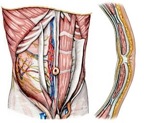
The anterior abdominal muscles include three very flat muscles and the M. rectus abdominis. The flat muscles radiate ventrally medially into the rectus sheath and insert on it via a flat tendon (aponeurosis). From outside to inside, the following muscles are found:
- M. obliquus externus abdominis: Dorsally, it originates from the thoracolumbar fascia as well as from the lower 7 ribs and extends as the anterior leaf of the rectus sheath to the median Linea alba as well as to the iliac crest of the pelvis. Its fibers run obliquely from cranial lateral to caudal medial.
- M. obliquus internus abdominis: It extends from the Linea alba to the iliac crest as well as to the anterior edge of the pubic bone. Its fibers also run obliquely from cranial medial to caudal lateral (in continuation of the M. obliquus externus abdominis of the opposite side). Both muscles thus form an oblique cross in the anterior abdominal wall. Above the Linea arcuata, it radiates into the anterior and posterior leaf of the rectus sheath, below the Linea arcuata only into the anterior leaf.
- M. transversus abdominis: Its fibers run from the thoracolumbar fascia or the lower costal cartilages and the pelvis to the Linea alba ventrally. It forms mainly the posterior leaf of the rectus sheath in the upper anterior abdominal wall. Below the Linea arcuata, it forms the anterior leaf together with the oblique abdominal muscles. The posterior wall of the 3 muscles is formed by the fascia transversalis.
- The M. rectus abdominis originates bilaterally from the cartilage of the 5th to 7th rib and inserts at the pubic bone near the pubic symphysis. Through intermediate tendons (intersectiones tendineae), the long muscle is divided into several bellies ("six-pack"). Inconsistently, the small M. pyramidalis is found caudally in front of the M. rectus abdominis, which tenses the Linea alba. The rectus sheath is thus a tendinous canal of the flat abdominal muscles, which, in addition to the M. rectus abdominis and M. pyramidalis, contains several vessels and nerves (Aa. and Vv. epigastricae superior and inferior, Nervi intercostales 5-12).
Function
For flexion and rotation of the trunk as well as for the abdominal press, the anterior abdominal wall is tensed in the described manner by the two oblique abdominal muscles (M. obliquus externus and internus abdominis, oblique cross) as well as the M. rectus abdominis and M. transversus abdominis (straight cross).
The M. cremaster is a detachment of the M. obliquus internus and M. transversus abdominis. It forms a muscular sheath around the spermatic cord and can pull the testicle cranially (cremaster reflex).
Deep Layer
The deep layer of the abdominal wall is formed by the fascia transversalis. It is found on the inside of the M. rectus abdominis and M. transversus abdominis as the innermost connective tissue layer (separated from the free abdominal cavity only by the peritoneum) and is fused with the Linea arcuata and the inguinal ligament. Laterally caudally, the anulus inguinalis profundus is located as the passage portal, which represents the entrance to the inguinal canal.
Dorsal Musculature
The main dorsal muscle of the abdominal wall is the M. quadratus lumborum, which extends below the M. transversus abdominis from the lowest rib and the costal processes of the lumbar vertebrae to the iliac crest.
Blood Supply and Innervation
The arterial supply is divided according to the listed layers of the abdominal wall:
- The superficial and middle layers are supplied by
→ the caudal Aa. intercostales posteriores (including the A. subcostalis),
→ the A. epigastrica superficialis,
→ the A. circumflexa ilium superficialis as well as
→ the A. pudenda externa. - The deep layer is supplied by
→ the Aa. lumbales,
→ the A. epigastrica inferior,
→ the A. circumflexa ilium profunda and
→ the A. iliolumbalis.
The venous blood of the abdominal wall drains via veins (predominantly → V. cava inferior) that are named corresponding to the arteries:
Via the V. epigastrica superficialis (→ V. saphena magna) as well as via the V. epigastrica inferior (→ V. iliaca externa). Only the venous blood reaches the Vena cava superior via the Vv. thoracoepigastricae as well as via the Vv. azygos and hemiazygos.
The innervation of the abdominal wall is provided by intercostal nerves and branches of the lumbar plexus:
- The lower Nn. intercostales (including the N. subcostalis) supply, as described, the M. obliquus externus abdominis and the M. rectus abdominis.
- From the lumbar plexus, the N. iliohypogastricus innervates all anterior abdominal wall muscles, the N. ilioinguinalis likewise all anterior abdominal wall muscles except the M. rectus abdominis, and the N. genitofemoralis supplies the M. transversus abdominis.
The Nn. iliohypogastricus and ilioinguinalis also run between the muscles they innervate to the skin of the anterior abdominal wall.
The lymphatic drainage of the anterior abdominal wall occurs above the umbilicus cranially (into the Nodi lymphatici axillares and parasternales), below the umbilicus caudally (into the Nodi lymphatici inguinales superficiales and iliaci). From the lateral abdominal wall, the lymph flows to the Nodi lymphatici lumbales.