Anatomy - Anesthesia examination, Pap smear, LEEP conization, cystoscopy, hysteroscopy, fractional curettage, rectoscopy

  1. The uterus

    The uterus

    The uterus, also known as the womb, is a hollow, muscular organ in the female body that plays an important role during pregnancy and childbirth.

    The uterus consists of:

    • the body (Corpus uteri), 
    • the isthmus uteri
    • the cervix (Cervix uteri) 

    The average length of the uterus is about 7.5 cm, the width 4 cm, and the thickness 2.5 cm. 

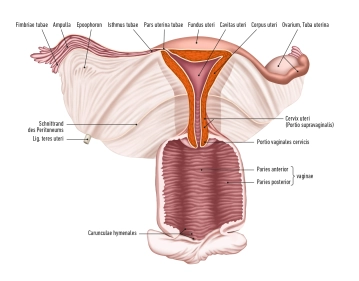
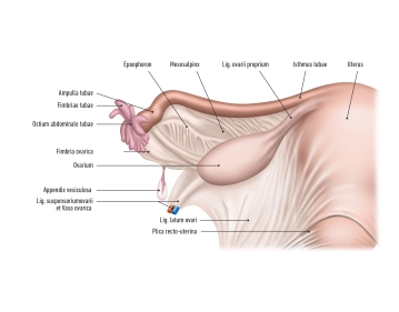
    The fundus: uteri represents the upper section of the uterus, located above the entry points of the fallopian tubes. Its lateral walls are connected with the broad ligament (Ligamentum latum uteri). At the lateral edges of the uterus, where the fallopian tubes enter the uterus, the tubal angle is formed. From the tubal angle, the proper ovarian ligament extends to the ovary, while the round ligament of the uterus extends to the inguinal canal.

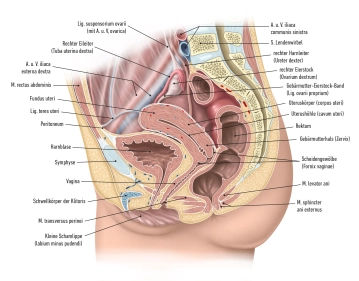
    Cervix uteri: The cervix extends with its conical lower third into the vagina. It is divided into the supravaginal portion of the cervix (Endocervix) and the vaginal portion of the cervix (Ectocervix or simply Portio). In the upper area of the vagina, the cervix uteri forms the anterior and posterior vaginal fornix (Fornix vaginae) between the portio and the vaginal wall.

    Portio vaginalis: At the end of the portio vaginalis is the external os, also known as the ostium uteri (Orificium externum uteri).

    The portio vaginalis normally has a reddish color and is covered with stratified, non-keratinized squamous epithelium, similar to the vagina. At the transition to the ostium uteri, this epithelium changes to the single-layered, columnar epithelium of the cervical canal (Canalis cervicis). Between puberty and menopause, the cylindrical epithelium of the cervical canal may migrate outward onto the portio (ectropion), leading to more intense redness. With a colposcopy, precancerous stages and cancer can be detected in this area.

  2. Hysteroscopy

    Hysteroscopy

    Cavum Uteri and Cycle-Dependent Changes in Hysteroscopy

    Cavum Uteri (Uterine Cavity):
    The cavum uteri is a triangular cavity extending from the inner surface of the fundus to the internal cervical os. During hysteroscopy, this structure provides a direct view of the endometrial surface and possible pathological changes.

    Tubal Ostia:

    • Tubal Ostia: The openings of the fallopian tubes (tubal ostia) are visible at the lateral upper corners of the cavum uteri. They appear as small, rounded openings.
    • Normal Form: Symmetrical and visible bilaterally in a normal uterine cavity.
    • Pathological Forms:
      • Closed or obstructed (indicating tubal pathology).
      • Abnormal positioning in uterine anomalies such as septate uterus.

    Corpus Uteri:

    • Normal Form: The uterine cavity has a smooth and even lining. In hysteroscopy, the walls appear homogeneous.
    • Pathological Forms:
      • Septate uterus: A partition partially or completely divides the uterine cavity.
      • Bicornuate uterus: Heart-shaped uterus with two horns.
      • Fibroids: Nodular growths protruding into the uterine cavity.
      • Polyps: Soft, benign tissue growths.

    Endometrial Changes:

    • Menstrual Phase: Fragmented and bloody.
    • Proliferative Phase: Smooth and even.
    • Secretory Phase: Spiral glands, highly vascularized.
    • Premenstrual Phase: Edematous and degenerative.

    Phases of the Menstrual Cycle and Endometrial Changes:

    1. Menstrual Phase (Day 1-4):

    • The endometrium appears thin and fragmented as the functional layer is shed.
    • Blood clots and shed tissue are visible.

    2. Proliferative Phase (Day 5-14):

    • Under the influence of estrogen, the endometrium thickens.
    • It shows a smooth, even surface with a thickness of about 1-3 mm.
    • Glands are elongated and straight, with clear, well-defined boundaries.

    3. Secretory Phase (Day 15-28):

    • Progesterone causes further changes in the endometrium, reaching a thickness of about 5-7 mm.
    • Glands become spiral and secrete a nutrient-rich secretion.
    • The stroma is edematous and shows significant vascular formation.

    4. Premenstrual Phase:

    • The endometrium is highly edematous and the glands are jagged.
    • There is increasing spiraling of the arterioles and beginning degeneration.
    Clinical Significance:

    Hysteroscopy can detect and assess various endometrial pathologies such as polyps, hyperplasia, or atrophy. The cycle-dependent changes of the endometrium help to accurately date the endometrium and identify any anomalies.

    Cervical Canal (Cervix):

    • Normal Findings:
      • Even and smooth canal.
      • Visible internal opening (internal os).
    • Pathological Findings:
      • Cervical stenosis: Narrowing of the cervical canal.
      • Polyps: Benign growths in the cervical canal.
      • Inflammations: Redness and swelling of the cervical mucosa.
      • Neoplasms: Noticeable growths that could indicate cancer.
    Clinical Significance: 

    Diagnosis of cervical pathologies such as polyps, stenosis, and inflammations. Assessment of cervical structure and identification of anomalies. Detection of precancerous changes through visual inspection of the mucosa and, if necessary, tissue sampling.

  3. The fallopian tube (Tuba uterina, Salpinx)

    The fallopian tube (Tuba uterina, Salpinx)

    The fallopian tube, also known as the Tuba uterina, is approximately 10-15 centimeters long and 2-5 millimeters thick. It runs intraperitoneally along the upper free edge of the broad ligament of the uterus (Ligamentum latum uteri), from the corner of the uterus to the ovary. In the mesosalpinx, the blood vessels and nerves run to the fallopian tube, additionally securing the fallopian tube to the Ligamentum latum.

    The fallopian tube itself can be divided into different sections:

    • Infundibulum tubae uterinae (+ Ostium abdominale tubae uterinae)
    • Fimbriae tubae uterinae,
    • Ampulla tubae uterinae
    • Isthmus tubae uterinae
    • Pars uterina.

    The blood supply to the Tuba uterina is provided by small branches (Rr. tubarii) from the arteries Arteria ovarica and Arteria uterina, which run in the mesosalpinx and anastomose with each other. Veins accompany the arteries and drain into the Vena ovarica and into the Plexus uterinus/uterovaginalis.

  4. Vagina

    Vagina

    Length and Structure: The vagina is a muscular and connective tissue organ with a length of about 8-11 cm. In its non-distended state, it shows a characteristic H-shaped structure in cross-section.

    Anatomical Orientation: Naturally, the vagina runs from bottom front to top back, adapted to the shape of the female pelvis.

    Vaginal Walls:

    • Paries anterior (Anterior Wall): This wall is shorter due to the presence of the portio vaginalis uteri, a part of the cervix that protrudes into the vagina.
    • Paries posterior (Posterior Wall): Longer than the anterior wall, it forms the larger posterior vaginal fornix (Fornix posterior).

    Vaginal Fornices (Fornix vaginae):

    • There are a total of four fornices: the posterior fornix, anterior fornix, and two lateral fornices, which form around the portio vaginalis of the uterus.

    Opening and Entrance:

    • Ostium vaginae: The opening of the vagina leads into the vestibule of the vagina, directly below the urogenital diaphragm.
    • Introitus vaginae: The entrance to the vagina, where the hymen or its remnants, the hymenal caruncles, are located.

    Vaginal Wall Features:

    • The wall of the vagina is soft in younger years and shows characteristic transverse folds, known as rugae vaginales, which contribute to flexibility and distensibility.

    Position in the Pelvis:

    • The vagina runs between the rectum and the bladder or urethra. Between these structures are the rectovaginal septum and the vesicovaginal or urethrovaginal septum, which serve as separating membranes.

    Features of the Posterior Vaginal Fornix:

    • The posterior fornix of the vagina forms the lower boundary of the rectouterine pouch, also known as the pouch of Douglas, and is covered by peritoneum.

    Vascular Supply:

    • The uterine artery reaches the cervix uteri near the lateral fornices of the vagina. At this anatomical site, the ureter crosses under the uterine artery, which is significant in surgical procedures.
  5. External genital organs

    External genital organs

    Mons pubis

    The mons pubis is a triangular elevation of connective tissue located above the symphysis. This elevation bears pubic hair in adult women.

    Labia majora and minora

    Labia majora:

    • The outer labia, also known as labia majora, contain subcutaneous fat tissue, forming them into raised skin folds.
    • They form a protective barrier for the vagina and enclose the labia minora.
    • The anterior and posterior connections of the labia majora are referred to as the commissura labiorum anterior et posterior.

    Labia minora:

    • The labia minora, also called labia minora, are thin, without fat tissue, but rich in sebaceous glands.
    • They surround the vestibule of the vagina.
    • Anteriorly, the labia minora form the frenulum clitoridis and the prepuce of the clitoris, posteriorly the frenulum labiorum.

    Vestibule of the vagina

    The vestibule of the vagina is laterally bounded by the labia minora, anteriorly by the frenulum clitoridis, and posteriorly by the frenulum labiorum. Within the vestibule are:

    • The vaginal opening (ostium vaginae)
    • The external urethral opening (ostium urethrae externum)
    • The ducts of the greater and lesser vestibular glands (glandulae vestibulares majores et minores)

    Glands of the vestibule

    Bartholin's glands (glandulae vestibulares majores):

    • These paired, pea-sized glands are located in the urogenital diaphragm.
    • The ducts open on the inner side of the labia minora, near the ostium vaginae.
    • They produce a secretion to lubricate the vaginal entrance.
    • If the ducts become blocked, Bartholin's cysts can form, and if these become inflamed, it is referred to as a Bartholin's abscess.

    Lesser vestibular glands:

    • These are collections of small glands that open into the vestibule of the vagina.

    Paraurethral glands (Skene's glands):

    • These glands open near the urethra and are also called paraurethral ducts.

    Hymen

    The hymen is a membrane that separates the external from the internal genital area.

    Vulva and erectile tissue

    Clitoris:

    • The clitoris consists of the crura clitoridis, the corpus clitoridis, and the glans clitoridis. It corresponds to the corpora cavernosa penis in males.

    Bulbus vestibuli:

    • The two bulbi vestibuli are located laterally to the vestibule of the vagina and correspond to the corpus spongiosum penis in males.
Rectum

Origin of the Rectum: Extension of the sigmoid colon, beginning at the upper edge of the third sacr

Activate now and continue learning straight away.

Single Access

Activation of this course for 3 days.

US$9.10  inclusive VAT

Most popular offer

webop - Savings Flex

Combine our learning modules flexibly and save up to 50%.

from US$4.20 / module

US$50.48/ yearly payment

price overview

gynecology

Unlock all courses in this module.

US$8.41 / month

US$101.00 / yearly payment