Start your free 3-day trial β€” no credit card required, full access included

Sigmoid resection, tubular, for diverticulitis, robotically assisted

Reading time readingtime 38:11 min.
Activate now
  1. Positioning

    507 Lagerung 1.jpeg

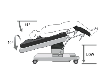
    Positioning is done in the lithotomy position on a large vacuum cushion. Positioning of both arms (caution: cotton wrapping when positioning with cloth sling). On the right side, the cushion supports the rib cage and the iliac crest, so that the patient's weight in right lateral position does not press on the arm. Positioning of the legs in padded “boots”/use of “swan-fins” for the legs, so that the legs can be moved separately and sterilely covered if necessary. Alternatively: Positioning of the legs in leg shells with fixation of the legs in these. Cotton wrapping of the knees and the proximal lower legs. The legs should be able to be flexed and extended via the OR table control

    Note: The positioning is of particular importance due to the docking of the patient to the robot's manipulator. Risk of injury to the abdominal wall if the patient slips.

    Caution: Vacuum cushions can have leaks. Check again before sterile covering.


  2. Trocar Positioning, Docking and TAP Block

    Video
    507_02.JPG
    Soundsettings

    Creation of a capnoperitoneum by inserting a Veress needle at Palmer's Point. The four 8mm robotic trocars are located on a straight line. The angle of the line corresponds to the connection of the intersection point of the left MCL / costal arch and the right femoral head. The line itself runs parallel 5-8 cm further to the right. Trocar I is located in the left epigastrium. Trocar IV two cm ventral to the right anterior superior iliac spine. Between the individual trocars there is a distance of ideally 8 cm (min 7 – max 10). The 12 mm assist trocar is located 4 cm cranial to Trocar 4 in the right anterior axillary line. The patient is positioned maximally to the right and moderately head down. The axis of the manipulator (laser marking) is aligned so that it points over the left anterior superior iliac spine to the camera trocar III. The arms are connected to the four 8mm robotic trocars (docked). Subsequently, the instruments are introduced under visual control and parked under the ventral abdominal wall. As the first step, diagnostic laparoscopy and inspection of the four quadrants are performed.

    Caution: The trocars must be positioned with the wide black ring at the level of the muscular abdominal wall (so-called remote center) to avoid injuring it during movement.

    Note: Ideally, the robotic trocars are inspected with the camera via the assist trocar when inserting the robotic instruments. This way, the position of all robotic trocars can be easily checked again before the start of the operation.

  3. Checklist up to Docking

    Checklist Xi Sigmoid Resection up to Docking

    • Stab incision left upper abdomen, insertion of the Veress needle
    • Pneumoperitoneum
    • Draw line and crosses for trocars 
    • Insertion of 4 Xi trocars 8 cm apart from each other
    • Insertion of the camera by hand after insertion of the first trocar and insertion of the others under vision
    • Camera trocar 12 mm with reducer sleeve on 3 or if necessary 4
    • Assistant trocar right mid-abdomen between 3 and 4
    • Positioning: >15° Trendelenburg, 10°Tilt right
    • Dock camera arm + insert camera 
    • Targeting
    • Dock 3 further arms
    • Arms always a fist's width apart
    • Check of the Remote Center
    • Burping
    • Insertion of the instruments and introduction into target anatomy (1: left cranial: Tip up, 2: bipolar forceps, 4: right caudal monopolar scissors/Vessel sealer, 3: Camera/Stapler)
    • Switch to the console
  4. Lateral Mobilization

    Video
    507_04 Toldt Linie.jpg
    Soundsettings

    Displacement of the small intestine to the right cranial  and, if necessary, hold it there by assistants. Grasping the sigmoid with the Tipup and pull medially so that the lateral structures tighten. Severing the post-inflammatory adhesions. Then lateral mobilization along the Toldt's line. In this way, the sigmoid and subsequently the descending colon are mobilized stepwise along the paracolic gutter in the avascular layer between the mesosigmoid and Gerota's fascia. The Gerota's fascia should remain intact. Identification of the left ureter dorsal to the intact Gerota's fascia.

    Note: The preparation is facilitated by the Trendelenburg position (“head down”) of the patient with simultaneous right tilt of the operating table.

    Caution: During the preparation, note not only the close proximity to the left ureter, but also to the gonadal vessels.


Mobilization of the left flexure

First, detachment of the omentum from the lateral abdominal wall with the Vessel-Sealer. Then, use

Activate now and continue learning straight away.

Single Access

Activation of this course for 3 days.

US$9.40  inclusive VAT

Most popular offer

webop - Savings Flex

Combine our learning modules flexibly and save up to 50%.

from US$4.37 / module

US$52.55/ yearly payment

price overview

robotics

Unlock all courses in this module.

US$8.75 / month

US$105.10 / yearly payment

to top