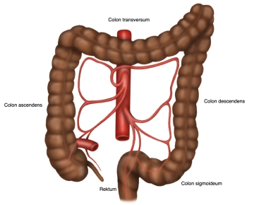
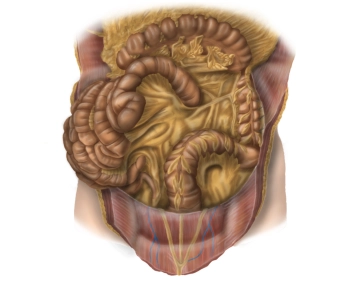
- The colon frames the small bowel loops along the inner abdominal wall and running below the liver and stomach. The position of the colon is intra- or secondarily retroperitoneal. The function consists mainly in the thickening of the chyme through the absorption of water. The length of the entire colon is on average 120-150 cm. The colon begins at the ileocecal valve and ends at the rectosigmoid junction, where it transitions into the rectum.
- The colon is divided into the sections:
- Cecum or caecum with the appendix
- Ascending colon
- Transverse colon
- Descending colon
- Sigmoid colon
- The rectum connects aborally to the sigmoid, begins about 15–18 cm above the anus and ends at the anorectal junction, where it transitions into the approximately 3–4 cm long anal canal.
-
Overview
![507 Anatomie Übersicht 2.jpg]()
-
Anatomy of the Descending Colon and Sigmoid Colon
![507 Anatomie 1.jpg]()
Descending Colon
Location: Secondary retroperitoneal.
Peritoneal Suspension: Fused with the dorsal abdominal wall.
Course: Extends from the left flexure (Flexura coli sinistra), which is fixed to the spleen via the phrenicocolic ligament, downward into the iliac fossa. It connects to the transverse colon and transitions distally into the sigmoid colon.
Length: Approximately 20–30 cm
Surgical Relevance: Firm fixation as a landmark for the proximal resection margin in sigmoid resection.
Sigmoid Colon (Sigma)
Location: Intraperitoneal.
Peritoneal Suspension: Sigmoid mesocolon.
Course: Begins in the left iliac fossa, typically describes an S-shaped loop and extends to the level of the 2nd–3rd sacral vertebra, where it transitions into the rectum.
Length: Variable (significantly longer in elongated sigma), on average about 35 cm
Surgical Relevance: Common location for diverticula; mesocolon mobilization for vascular preparation and anastomosis planning.
-
Macroscopy
![507 Makroskopie Colon.jpg]()
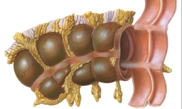
- The longitudinal muscle layers of the colon form three band-like muscle strips that are visible from the outside and are called taeniae. They are distinguished as:
- Taenia mesocolica: lying towards the mesentery
- Taenia libera: free on the surface, facing the abdominal wall
- Taenia omentalis: connected to the greater omentum
- Appendices epiploicae are fat appendages from the subserous layer in the area of the free taeniae.
- Plicae semilunares are indentations of all wall layers towards the intraluminal side, whereas haustra are the intervening outward bulges.
- For the sigmoid resection, the following intestinal segments from oral to aboral are relevant:
- left flexure
- descending colon
- sigmoid colon
- upper rectum
- The longitudinal muscle layers of the colon form three band-like muscle strips that are visible from the outside and are called taeniae. They are distinguished as:
Vascular and Nerve Supply, Lymphatic Drainage
Arterial SupplyThe left hemicolon, the sigmoid colon up to the upper rectum are supplied by the inf
Arterial SupplyThe left hemicolon, the sigmoid colon up to the upper rectum are supplied by the inf
Activate now and continue learning straight away.
Single Access
Activation of this course for 3 days.
US$9.30
inclusive VAT
Most popular offer
webop - Savings Flex
Combine our learning modules flexibly and save up to 50%.
from US$4.32 / module
US$51.88/ yearly payment
robotics
Unlock all courses in this module.
US$8.64
/ month
US$103.80 / yearly payment
Webop is committed to education. That's why we offer all our content at a fair student rate.