Start your free 3-day trial — no credit card required, full access included

Anatomy - Aortobifemoral bypass for peripheral arterial disease Fontaine stage IIb–III

  1. Abdominal aorta

    Abdominal aorta
    Zum Vergrößern bitte anklicken

    Origin

    • From the thoracic aorta after its passage through the aortic hiatus in the diaphragm at the level of T12

    Course

    • Retroperitoneal
    • Left to the median plane and anterior to the spine
    • Bifurcation into the common iliac arteries (bifurcatio aortae) at the level of the umbilicus / L4

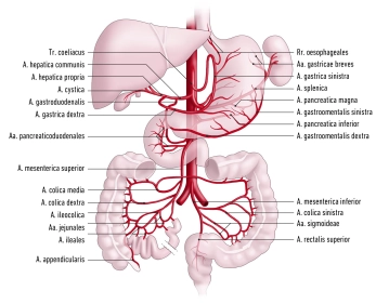
    Branches from superior to inferior

    • Inferior phrenic artery, left and right
    • Celiac trunk
    • Middle suprarenal artery, left and right
    • Superior mesenteric artery
    • Renal artery, left and right
    • Ovarian / testicular artery, left and right
    • Lumbar arteries
    • Inferior mesenteric artery
    • Median sacral artery

    Distribution

    • Paired branches: Abdominal wall, paired retroperitoneal organs, gonads
    • Unpaired branches: Spleen, unpaired intestinal organs
  2. Paired branches of the abdominal aorta

    Main branches

    Course

    Branches/rami and distribution

    Inferior phrenic artery

    • Travels craniad along the underside of the diaphragm towards the diaphragmatic dome
    • Network-like division into several terminal branches

    Superior suprarenal artery: Suprarenal gland

    Terminal branches: Underside of the diaphragm

    Middle suprarenal artery

    • Travels laterad to the suprarenal glands

    Terminal branches: Suprarenal glands

    Renal artery

    • Arises at the level of L1–2
    • Right renal artery: Travels posterior to the vena cava and pancreatic head to the right kidney
    • Left renal artery: Travels posterior to the body of the pancreas to the left kidney

    Inferior suprarenal artery: Suprarenal gland

    Ovarian artery 

    Testicular artery

    • Arises from the anterior wall of the aorta inferior to the renal arteries / L2
    • Travels across the psoas muscle, ureter, and external iliac artery to the suspensory ligament of ovary / deep inguinal ring and testis

    Ovarian arteries: Ovaries, parts of the uterine tubes

    Testicular arteries: Testes, epididymides

    Lumbar arteries (4 bilat.)

    • Travels lateroposteriad to the psoas muscle across L1–4
    • Anastomose with the arteries of the anterior abdominal wall (superior and inferior epigastric arteries; liliolumbar artery; deep circumflex iliac artery)

    Spinal rami: Spinal canal

    Terminal branches: Abdominal wall, back muscles

  3. Unpaired branches of the abdominal aorta

    All unpaired branches of the abdominal aorta terminate in anastomoses maintaining organ perfusion via collateral blood supply:

    • Celiac trunk -> gastroduodenal artery -> superior mesenteric artery
    • Superior mesenteric artery -> middle and left colic artery -> inferior mesenteric artery (anastomosis of Riolan)
    • Inferior mesenteric artery -> superior and middle rectal artery -> internal iliac artery


    1. Celiac trunk
    • Arises at the level of T12
    • Divides into the splenic, left gastric, and common hepatic arteries (biggest caliber of the celiac trunk)

    Main branches

    Course

    Branches/rami and their course 

    Distribution

    Splenic artery

    • Along the superior aspect of the pancreas
    • Enters the splenorenal ligament and spleen

    Pancreatic rami:

    • Posterior to the pancreas

    Dorsal pancreatic artery:

    • Posterior to the pancreas
    • Anastomoses with the great pancreatic artery to the inferior pancreatic artery

    Dorsal pancreatic artery:

    • Posteroinferior along the pancreatic tail
    • Anastomoses with the dorsal pancreatic artery to the inferior pancreatic artery

    Pancreas

    Posterior gastric artery:

    • Travels in the phrenicosplenic ligament superior to the gastric fundus

    Stomach (posterior wall of fundus)

    Short gastric arteries:

    • Travel in the gastrosplenic ligament to the gastric fundus

    Stomach (fundus and greater curvature)

    Left gastro-omental artery:

    • Travels in the gastrocolic ligament to the greater curvature
    • Anastomoses with the right gastro-omental artery

    Stomach (fundus and greater curvature)

    Left gastric artery

    • Travels to the lesser gastric curvature
    • Anastomoses with the right gastric artery

    Direct branches

    Stomach (lesser curvature)

    Esophageal branches

    Esophagus

    Common hepatic artery

    • Travels to the hepatic portal on the right
    • Bifurcates into the hepatic artery proper and gastroduodenal artery

    Proper hepatic artery

    • Main branch of the common hepatic artery
    • Arises posterior to the pylorus
    • Travels in the hepatoduodenal ligament together with the hepatic portal vein and common bile duct to the hepatic portal
    • Divides into 3 branches :

    1. Right gastric artery:

    • Travels in the minor omentum to the lesser gastric curvature
    • Anastomoses with the left gastric artery

    Stomach (lesser curvature on right)

    2. Right hepatic artery

    Liver( right part)

    Gallbladder (cystic artery)

    Parts of caudate lobe

    3. Left hepatic artery

    Liver( left part)

    Parts of caudate lobe

    Gastroduodenal artery

    • Travels posterior to the lesser omentum to the pancreas
    • Divides into 2 branches:

    1. Right gastro-omental artery:

    • Runs to the greater gastric curvature
    • Anastomoses with the left gastro- omental artery

    Pancreas, duodenum, stomach

    2. Anterior and posterior superior pancreaticoduodenal artery

    • together with branches of the inferior pancreaticoduodenal artery form a vascular plexus around the pancreatic head


    2. Superior mesenteric artery
    • Arises from the aorta at the level of L1–2
    • Travels anterocaudad
    • Branches: Inferior pancreaticoduodenal artery; jejunal and ileal arteries; ileocolic arteries; right colic artery; middle colic artery

    Main branches

    Course

    Branches/Rami

    Distribution

    Inferior pancreaticoduodenal artery

    • Travels from the horizontal part of duodenum craniad to the pancreatic head

    Anterior/posterior ramus

    • Anastomoses with the anterior/posterior pancreaticoduodenal artery respectively

    Pancreatic head

    Jejunal and ileal arteries

    • Travel in the root of the mesentery to the jejunum and ileum
    • Anastomose with each other to form an arcade

    Rectal arteries

    Jejunum, ileum

    Ileocolic artery

    • Travels in the mesentery to the ileocecal region

    Anterior/posterior cecal artery

    Cecum

    Appendicular artery (variant: arises from the posterior cecal artery)

    Appendix

    Right colic artery

    • Travels in the ascending mesocolon to the ascending colon

    Bifurcates into 2 branches which anastomose with the middle colic and ileocolic artery respectively

    Ascending colon

    Middle colic artery

    • Travels in the transverse mesocolon to the transverse colon

    Bifurcates into 2 branches: Right branch anastomoses with the right colic artery (right colic flexure). Left branch anastomoses with the left colic artery (left colic flexure)

    Transverse colon

    3. Inferior mesenteric artery
    • Arises from the aorta at the level of L3
    • Supplies the colon downstream of the left colic flexure via left colic artery, sigmoid arteries, and superior rectal artery

    Main branches

    Course

    Branches/Rami

    Supplies

    Left colic artery

    • Travels in the descending mesocolon to the descending colon

    Bifurcates into 2 branches which anastomose with the middle colic and sigmoid arteries

    Descending colon

    Sigmoid arteries

    • 2–4 arteries anastomosing to an arcade in the sigmoid mesocolon
    • Anastomose with the left colic and superior rectal arteries

    several small branches to the sigmoid

    Sigmoid

    Superior rectal artery

    • Approaches the rectum from posterior
    • Anastomoses with the middle rectal artery

    several small branches to the rectum

    Upper rectum

  4. Arterial blood supply of the lesser pelvis

    Arterial blood supply of the lesser pelvis
    Zum Vergrößern bitte klicken
    • At the aortic bifurcation (at about L4), the abdominal aorta divides into both common iliac arteries
    • each common iliac artery in turn bifurcates into the internal and external iliac arteries
    • With its visceral branches, the internal iliac artery mainly supplies the pelvic organs, while with its parietal branches contribute to the blood supply of the lower limbs.
    • The external iliac artery contributes to the blood supply of the pelvis and becomes the femoral artery after passing through the vascular lacuna
    1. Internal iliac artery

    Origin

    • arises from the common iliac artery directly inferior to the aortic bifurcation

    Course

    • passes caudad over the superior margin of the sciatic foramen

    Relationships

    • Posterior: Internal iliac vein, piriform muscle, lumbosacral plexus
    • Anterior: Ureter
    • Lateral: External iliac vein, greater psoas muscle
    • Inferior:  Obturator nerve

    Branches/Rami

    Visceral branches:

    • Umbilical artery
    • Inferior vesical artery
    • Middle rectal artery
    • In women: Uterine artery

    Parietal branches

    • Iliolumbar artery
    • Lateral sacral arteries
    • Superior gluteal artery
    • Inferior gluteal artery
    • Obturator artery
    • Internal pudendal artery

    Distribution

    • Pelvic organs, gluteal muscles
    1.1 Visceral branches of the internal iliac artery

    Main branches

    Course and branches

    Distribution

    Umbilical artery

    • Postnatal obliteration to median umbilical ligament
    • Remaining branches: Superior vesical arteries, in men: Artery of ductus deferens
    • Body of bladder
    • In men: Ductus deferens

    Inferior vesical artery

    • Divides into small branches at the fundus of the bladder
    • Bladder
    • In men: Prostate, seminal vesicles

    Middle rectal artery

    • Divides at the level of the middle rectum
    • Anastomoses with the inferior and superior rectal arteries
    • Rectal ampulla
    • In women: Vagina
    • In men: Prostate

    Uterine artery (women)

    • Crosses the ureter
    • Tortuous course in the broad ligament of uterus
    • Gives off numerous small branches
    • Ovary
    • Uterine tube
    • Proximal vagina
    • Uterus
    1.2 parietal branches of the internal iliac artery

    Main branches

    Course and branches

    Distribution

    Iliolumbar artery

    • Travels obliquely craniad to the iliopsoas muscle
    • Major psoas muscle
    • Iliac muscle

    Lateral sacral arteries

    • Superior ramus: runs mediad through the first or second anterior sacral foramen and leaves via the posterior sacral foramen
    • Inferior ramus: runs across the piriform muscle and medial to the sacral foramina caudad on the sacrum
    • Sacral canal
    • Piriform muscle
    • Muscles and skin posterior to the sacrum

    Superior gluteal artery

    • Travels together with the superior gluteal nerve through the suprapiriform foramen and with branches to the gluteus maximus muscle (superficial branch) and gluteus medius muscle (deep branch)
    • Gluteus maximus/medius/minimus muscles
    • Piriform muscle
    • Tensor muscle of fascia lata
    • Skin over sacrum

    Inferior gluteal artery

    • Travels caudad on the piriform muscle through the infrapiriform foramen
    • Then passes together with the sciatic and cutaneous femoral nerves through the gluteus maximus muscle
    • Gluteus maximus/medius/minimus muscles
    • Piriform muscle
    • Quadriceps femoris muscle
    • Skin of the inferior buttocks and proximal thigh

    Obturator artery

    • Travels anterocaudad along the lateral pelvic wall
    • Branches to the iliac muscle and bone
    • after passing through the obturator canal, further branches originate, including the . pubic ramus (anastomoses with the obturator ramus of the inferior epigastric artery to the corona mortis), acetabular ramus (-> ligament of head of femur (lig. capitis femoris), anterior ramus (anastomoses with the medial circumflex femoral artery), and posterior ramus
    • Iliac bone
    • Iliac muscle
    • Adductor muscles of the thigh
    • Pubic bone
    • Lateral thigh muscles
    • Head of femur

    Internal pudendal artery

    • Travels anterior to the sacral plexus to the infrapiriform foramen
    • Reenters the lesser pelvis through the lesser sciatic foramen, passes along the pudendal canal and terminates in numerous rami in the ischioanal (ischiorectal) fossa
    • Distal third of rectum
    • Anal canal
    • Urethra
    • Scrotal skin / labia
    • Bulb of penis /vestibule of vagina
    • Dorsum of penis / prepuce of clitoris
    2. External iliac artery

    Origin

    • Arises from the common iliac artery inferior to the internal iliac artery

    Course

    • Travels along the greater psoas muscle to the inguinal ligament and becomes the femoral artery once it passes through the vascular lacuna

    Relationships

    • The external iliac artery passes posterior to the ureter

    Branches/Rami

    • Deep circumflex artery travels to the superior iliac spine and anastomoses with the iliolumbar and superior gluteal arteries
    • Inferior epigastric artery  runs craniad medial to the inguinal ring and between the transverse abdominal and rectus abdominis muscles, other branches include the cremasteric artery / artery of round ligament of uterus

    Distribution

    • Iliac bone. Inferior abdominal wall