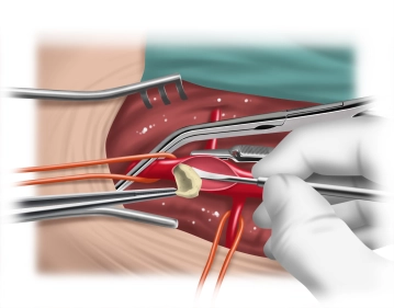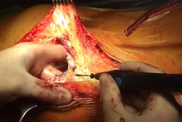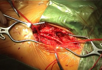
Longitudinal skin incision about 1 cm lateral to the femoral artery and transection of the subcutis with meticulous haemostasis. While sparing the lymphatics, divide the lateral lymph collectors immediately caudal to the groin between clamps and secure them with transfixion ligatures. Dissect and loop the common femoral artery, superficial femoral artery, and deep femoral artery. Palpate the vessel wall (quality, calcification).
Tips:
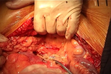
1. The first step is to expose the groin so that the abdominal cavity remains open only for a short time. As a result, the patient cools down less quickly, which improves the postoperative phase and allows for faster extubation.
2. Access lateral to the femoral artery allows a staggered closure of the groin. This better secures the vascular reconstruction, and superficial wound healing disorders, such as wound margin necrosis, do not necessarily result in a deep infection.
3. Especially in Fontaine stage IV PAOD, the lymph collectors and lymph nodes are often markedly enlarged and represent a potential source of infection for the vascular reconstruction. Careful instrumental dissection is therefore imperative, with no crude manipulation of the wound with the fingers!
4. The medial circumflex femoral artery lies mediodorsally (femoral artery -> deep femoral artery -> medial circumflex femoral artery). Its location and caliber vary greatly and it is often of large caliber. It must be looped separately and clamped off later, since injuries to this vessel can lead to bleeding that is difficult to manage.
5. In Fontaine stage IV PAOD, as well as in repeat procedures, pronounced adhesions between the femoral artery and the deep femoral vein are usually present. In these cases, time-consuming dissection is to be expected.