Start your free 3-day trial — no credit card required, full access included

Perioperative management - Aortofemoral bifurcation bypass (bifurcated graft) for peripheral arterial occlusive disease stage IIb–III

  1. Indications

    Depending on the length of the local occlusion/stenosis, the TASC criteria allow therapeutic treatment options to be derived. The length of the stenosis and the regions where it is located determine the type of management: endovascular or open vascular surgery.

    TASC criteria for aortoiliac arterial occlusions

    Lesion type

    Morphology

    Recommended treatment

    A

    Focal stenoses of the common iliac artery or external iliac artery <3 cm, unilateral or bilateral

    Endovascular

    B

    Focal stenoses 3–10 cm long and/or unilateral occlusion of the common iliac artery

    Endovascular

    C

    Bilateral stenoses of the common iliac artery, 5–10 cm, or unilateral complete occlusion of the external iliac artery or bilateral occlusions of the common iliac artery

    Open reconstruction

    D

    Diffuse stenotic changes of the entire iliac axis, or unilateral occlusion of the common and external iliac artery, or bilateral occlusions of the external iliac artery

    Open reconstruction

    Video example:

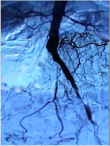
    Präoperatives Angiogramm
    Preoperative angiogram

    Clinically the patient is classified as Fontaine PAOD IIb–III and the preoperative angiogram demonstrates: occlusion of the right common iliac artery and external iliac artery, subtotal stenosis of the left external iliac artery including bilateral stenosis of the femoral bifurcation.

    -> TASC D, thus recommendation for open revascularisation

    PAOD classification according to Fontaine stages and Rutherford categories


    Fontaine stage

    Clinical symptoms

    Rutherford category

    Grade

    Clinical symptoms

    I

    Asymptomatic

    0

    0

    Asymptomatic

    IIa

    Distance > 200 m

    1

    I

    Mild intermittent claudication

    IIb

    Distance < 200 m

    2

    I

    Moderate intermittent claudication

    3

    I

    Severe intermittent claudication

    III

    Ischaemic rest pain

    4

    II

    Ischaemic rest pain

    IV

    Ulceration, gangrene

    5

    III

    Small-area necrosis

    6

    III

    Large-area necrosis

  2. Contraindications

    • Serious cardiopulmonary risks (e.g. NYHA IV, COPD GOLD stage IV)
    • Acute or chronic inflammatory abdominal disease (e.g. florid ulcerative colitis, recurrent sigmoid diverticulitis)
    • History of multiple extensive abdominal procedures ("hostile abdomen")
    • Cirrhosis
    • Advanced malignancy
  3. Preoperative diagnostic work-up

    Medical history

    • Claudication
    • Walking distance
    • Risk factors -> nicotine; arterial hypertension; CHD; heart failure; diabetes mellitus; manifest renal failure with/without dialysis dependency; coagulation disorders

    Inspection

    • Skin changes
    • Muscular abnormalities
    • Orthopaedic deformities
    • Skin colour
    • Hair
    • Trophic changes
    • Swelling; oedema; mycosis; phlegmon; leg ulcers etc.

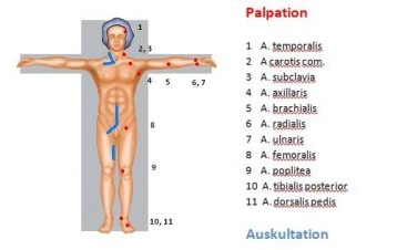
    Palpation with contralateral comparison

    • Arterial pulse examination
    • Skin temperature

    Arterial auscultation of the extremities with contralateral comparison

    Palpation-Auskultation
    Palpation–auscultation

    Ankle-Brachial Index (ABI)

    • ABI = systolic BP of the posterior tibial artery / systolic BP of the brachial artery

    ABI value

    Severity of PAOD

    > 1.3

    Falsely high values (suspected Mönckeberg medial sclerosis, e.g. in diabetes mellitus)

    > 0.9

    Normal finding

    0.75 - 0.9

    Mild PAOD

    0.5 - 0.75

    Moderate PAOD

    < 0.5

    Severe PAOD

    • An ABI of < 0.9 is considered proof of the presence of significant PAOD.
    • Noninvasive Doppler ultrasound measurement of the occlusion pressure to determine the Ankle-Brachial Index (ABI) is an adequate test for the presence of PAOD.
    • For the diagnosis of PAOD, the ABI with the lowest ankle artery pressure is used.
    • A pathological ankle-brachial index is an independent risk indicator for increased cardiovascular morbidity and mortality.

    Colour-coded duplex sonography

    • Carotid arteries, abdominal aorta, arteries of the extremities
    • Localisation of stenoses and occlusions in almost all vascular regions except the thoracic region
    • Quantification of the degree of stenosis and assessment of plaque morphology possible
    • Sensitivity and specificity about 90%.
    • Well suited as a screening method

    CT angiography

    • Multi-slice computed tomography (MS-CT) using non-ionic contrast medium
    • Broad range of indications: traumatic vascular lesion (especially trunk); vascular dissection/rupture; aneurysm; arterial thrombosis/embolism; portal vein/mesenteric vein thrombosis; pulmonary artery embolism; PAOD; vascular tumours
    • Pros: quickly performed; detection of relevant concomitant disease; imaging of peripheral arteries; sensitivity and specificity each about 90%
    • Cons: exposure to radiation and contrast media; allergies (about 3%); no functional assessment

    Cardiac check-up

    • Resting ECG
    • Exercise ECG
    • Echocardiography

    Chest radiograph

    Spirometry if required

    Clinical chemistry

    • FBC
    • Electrolytes
    • Coagulation
    • Renal function
    • Liver function
    • Blood lipids
    • Blood group
  4. Special preparation

    • Enema the evening before surgery
    • Hair trimmed in the surgical field
    • Packed RBCs ordered
    • Indwelling urinary (Foley) catheter
    • Perioperative prophylactic antibiotics 30 minutes before surgery (see German KRINKO and Robert-Koch-Institut recommendations)
  5. Informed consent

    General surgical risks

    • Major bleeding; blood transfusions; hepatitis/HIV transmission from allogeneic blood units
    • Allergy/incompatibility
    • Wound infection
    • Thrombosis/embolism
    • Injury to skin, vessels and nerves, e.g. due to positioning
    • Keloids
    • Incisional hernia

    Specific surgical risks

    • Thrombosis of the vascular graft and possibly adjacent vascular segments, possibly leg ischaemia, amputation
    • Graft infection with bleeding from suture lines; sepsis; leg ischaemia; amputation
    • Injury to adjacent organs such as ureter, bladder, spleen, liver; intestinal ischaemia -> resection, stoma formation
    • Paraplegia in the case of a low-origin arteria radicularis magna
    • Nerve lesions -> paraesthesia; pain; paralysis of the abdominal wall and thigh muscles
    • Peritoneal adhesions -> chronic pain, mechanical ileus
    • Lymphatic fistula
    • Secondary bleeding
    • Impotence
    • Anastomotic/graft aneurysm
    • Impaired renal function due to intraoperative angiography
Anaesthesia

General anaesthesia ... - Operations in general, visceral and transplant surgery, vascular surgery

Activate now and continue learning straight away.

Single Access

Activation of this course for 3 days.

US$9.30  inclusive VAT

Most popular offer

webop - Savings Flex

Combine our learning modules flexibly and save up to 50%.

from US$4.30 / module

US$51.61/ yearly payment

price overview

vascular surgery

Unlock all courses in this module.

US$8.60 / month

US$103.20 / yearly payment

to top