Start your free 3-day trial โ€” no credit card required, full access included

Endovascular bifurcation prosthesis for infrarenal AAA with simultaneous aneurysm of the right common iliac artery (EVAR with iliac side branch)

Reading time readingtime 32:04 min.
Activate now
  1. Principle of the Endovascular Procedure

    Video
    Principle of the Endovascular Procedure
    Soundsettings

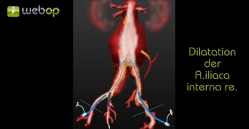
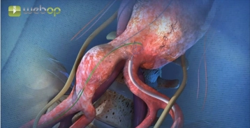
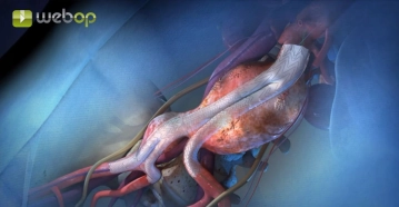
    After puncturing both groin arteries, two guidewires are first introduced from the right via a sheath. From the left, a Dormia basket is introduced through a long sheath. This is used to grasp one of the stiff wires and pull it from right to left in the groin over the sheath, so that the wire can be tensioned from both groins in order to position the endoluminal prostheses in the subsequent steps. Now the iliac prosthesis component with a side arm for the internal iliac artery is introduced from the right using a delivery system, partially deployed, and positioned. Then probing is performed with a delivery system from the left to advance the extension into the side arm up to the internal iliac artery and deploy it. Next, the iliac prosthesis component is completely released. In a further step, after probing the aorta with a stiff wire from the left femoral side, a delivery system with the Y-prosthesis main body is introduced and deployed. Finally, the main body and the left-sided iliac prosthesis module are connected with a further tubular prosthesis segment.

    In our case example, various steps were modified because it was necessary to dilate stenoses of the right common and internal iliac arteries. In addition, a further extension was introduced on the left iliac side for surgical-tactical reasons.

    (Copyright (c) 2015 by W.L. Gore and Associates GmbH. Used with permission.)
  2. Bilateral inguinal access

    Video
    Bilateral inguinal access
    Soundsettings

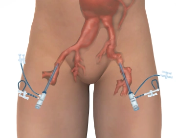
    Longitudinal incision in the region of both groins approximately 1 cm lateral to the palpable inguinal vessels, exposure and looping of the common femoral artery. Puncture of the femoral arteries on both sides using the Seldinger technique and insertion of suitable sheaths (e.g., 6F). Administration of 5,000 IU of heparin on each side. Insertion of a pigtail catheter for angiography from the right. On the right, the stenosed external iliac artery is predilated with an 8 mm balloon.

    Tips:

    1. Access lateral to the femoral artery spares the lymphatic collectors and later enables a curtain closure with the aim of secure wound healing.

    2. Note the medial circumflex femoral artery, which sometimes lies hidden dorsally. Back-bleeding from this artery can be very intense.

    3. All subsequent manipulations with guidewires and prosthesis segments should always be performed under fluoroscopic control to avoid perforations and malpositioning.

  3. Crossover catheterisation of the right common iliac artery, dilatation of the right internal iliac artery origin

    Video
    Crossover catheterisation of the right common iliac artery, dilatation of the right internal iliac artery origin
    Soundsettings

    Insertion of a guidewire (e.g. 0.035'' Terumo®) transfemorally from the left under fluoroscopic control. Catheterisation of the right common iliac artery with a diagnostic or guiding catheter (SIMS 1, Hockey Stick or others) in crossover technique, then catheterisation of the right internal iliac artery. In this case the right internal iliac artery origin is severely stenosed and is predilated with a 6 mm balloon. This is done in kissing balloon technique (internal and external/common iliac artery).

  4. Insertion and prepositioning of the iliac branch component

    Video
    Insertion and prepositioning of the iliac branch component
    Soundsettings

    First, using a snare catheter from the left, a guidewire introduced from the right is captured and led out to the left, so that the wire ends can be tensioned on both sides to allow good positioning of even a bulky endoluminal prosthesis. This wire was introduced alongside the first right-sided guidewire. The iliac branch component is prepared and flushed with heparinised saline solution. From the right, the iliac component can then be advanced and placed.

    Tip:

    The branch component must be placed in such a way that the lumen of the left common iliac artery is not obstructed. Furthermore, the branch component should come to lie a few millimetres before the origin of the right internal iliac artery to ensure catheterisation.

Partial deployment of the iliac branch component

The iliac part of the prosthesis is slowly partially deployed and carefully brought into position,

Activate now and continue learning straight away.

Single Access

Activation of this course for 3 days.

US$9.30  inclusive VAT

Most popular offer

webop - Savings Flex

Combine our learning modules flexibly and save up to 50%.

from US$4.30 / module

US$51.61/ yearly payment

price overview

vascular surgery

Unlock all courses in this module.

US$8.60 / month

US$103.20 / yearly payment

to top