Start your free 3-day trial — no credit card required, full access included

Anatomy - Endovascular bifurcation prosthesis for infrarenal AAA with simultaneous aneurysm of the right common iliac artery (EVAR with iliac side branch)

  1. Abdominal aorta

    Abdominal aorta
    Zum Vergrößern bitte anklicken

    Origin

    • from the thoracic aorta after its passage through the aortic hiatus of the diaphragm at the level of T12

    Course

    • retroperitoneal
    • left of the median plane, ventral to the vertebral column
    • at the level of the umbilicus/L4, division (aortic bifurcation) into the common iliac arteries

    Branches from cranial to caudal

    • Inferior phrenic arteries
    • Celiac trunk
    • Middle suprarenal artery, right and left
    • Superior mesenteric artery
    • Right or left renal artery
    • Ovarian artery - or testicular artery, right and left
    • Lumbar arteries
    • Inferior mesenteric artery
    • Median sacral artery

    Supply area

    • paired branches: abdominal wall, paired retroperitoneal organs (kidneys, adrenal glands), gonads
    • unpaired branches: spleen, liver, pancreas, unpaired digestive organs
  2. Paired branches of the abdominal aorta

    Main branches

    Course

    Branches and their supply areas

    Inferior phrenic artery

    • extends cranially on the underside of the diaphragm towards the diaphragmatic domes
    • network-like division into several terminal branches

    Superior suprarenal artery: Adrenal gland

    Terminal branches: Underside of the diaphragm

    Middle suprarenal artery

    • extends laterally to the adrenal glands

    Terminal branches: Adrenal glands

    Renal artery

    • Origin approx. at the level of L1 - 2
    • Right renal artery: runs dorsal to the inferior vena cava and pancreatic head to the right kidney
    • Left renal artery: runs dorsal to the pancreatic body to the left kidney

    Inferior suprarenal artery: Adrenal gland

    Ovarian artery,

    Testicular artery

    • Origin below the renal arteries/L2 from the anterior aspect of the aorta
    • run over the psoas muscle, ureter, and external iliac arteries to the suspensory ligament of the ovary or to the deep inguinal ring of the inguinal canal and the testis

    Ovarian arteries: Ovaries, parts of the uterine tube,

    Testicular arteries: Testes, epididymides

    Lumbar arteries (4 on each side)

    • extend laterally behind the psoas muscle over L1 – 4
    • Anastomoses with the arteries of the anterior abdominal wall (superior and inferior epigastric arteries, iliolumbar artery, deep circumflex iliac artery)

    Spinal branches: Spinal canal

    Terminal branches: Abdominal wall, back musculature

  3. Unpaired branches of the abdominal aorta

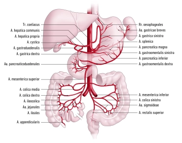
    • All unpaired branches of the abdominal aorta form anastomoses that maintain organ perfusion via a collateral supply:
      • Coeliac trunk → gastroduodenal artery → superior mesenteric artery
      • Superior mesenteric artery → middle and left colic artery → inferior mesenteric artery (“Riolan anastomosis”, “Drummond arcade”, rarely the anastomosis according to William and Klopp)
      • Inferior mesenteric artery → superior and middle rectal artery → internal iliac artery
    1. Coeliac trunk
    • Origin at the level of T12
    • divides into the splenic artery, left gastric artery, and common hepatic artery (the largest-calibre branch of the trunk)

    Main branches

    Course

    Branches and their course

    Supply area

    Splenic artery

    • Along the upper border of the pancreas
    • Entry into the splenorenal ligament and spleen

    Pancreatic branches:

    • Dorsal to the pancreas

    Dorsal pancreatic artery:

    • Dorsal to the pancreas
    • Anastomoses with the great pancreatic artery to form the inferior pancreatic artery

    Great pancreatic artery:

    • Dorsal to the pancreatic tail, running caudally
    • Anastomoses with the dorsal pancreatic artery to form the inferior pancreatic artery

    Pancreas

    Posterior gastric artery:

    • Runs in the phrenicosplenic ligament cranially to the gastric fundus

    Stomach (posterior surface of the fundus)

    Short gastric arteries:

    • Run in the gastrosplenic ligament to the gastric fundus

    Stomach (fundus and greater curvature)

    Left gastro-omental artery:

    • Runs in the gastrocolic ligament to the greater curvature
    • Anastomoses with right gastro-omental artery

    Stomach (fundus and greater curvature)

    Left gastric artery

    • Runs to lesser gastric curvature
    • Anastomoses with right gastric artery

    Direct branches

    Stomach (lesser curvature)

    Oesophageal branches

    Oesophagus

    Common hepatic artery

    • Runs to the right towards the hepatic portal
    • Divides into the proper hepatic artery and gastroduodenal artery

    Proper hepatic artery

    • Main branch of the common hepatic artery
    • Arises behind the pylorus
    • Runs in the hepatoduodenal ligament together with the hepatic portal vein and common bile duct to the hepatic portal
    • Gives off 3 branches:

    1. Right gastric artery:

    • Runs in the lesser omentum to the lesser gastric curvature
    • Anastomoses with left gastric artery

    Stomach (lesser curvature, right)

    2. Right hepatic artery

    Liver (right part)

    Gallbladder (cystic artery)

    Partially caudate lobe

    3. Left hepatic artery

    Liver (left part)

    Partially caudate lobe

    Gastroduodenal artery

    • Runs dorsal to the lesser omentum to the pancreas
    • Divides into 2 branches:

    1. Right gastro-omental artery:

    • Runs to the greater curvature
    • Anastomoses with left gastro-omental artery

    Pancreas, duodenum, stomach

    2. Anterior and posterior superior pancreaticoduodenal artery:

    • Together with branches of the inferior pancreaticoduodenal artery, form a vascular plexus around the pancreatic head
    2. Superior mesenteric artery
    • Arises from the aorta at the level of L1-2
    • Runs anteroinferiorly
    • Branches: inferior pancreaticoduodenal artery, jejunal arteries and ileal arteries, ileocolic artery, right colic artery, middle colic artery

    Main branches

    Course

    Branches

    Supply area

    Inferior pancreaticoduodenal artery

    • Runs in the region of the horizontal part of the duodenum cranially to the pancreatic head

    Anterior and posterior branches:

    • Anastomose with the anterior and posterior pancreaticoduodenal artery

    Pancreatic head

    Jejunal arteries and ileal arteries

    • Run in the mesenteric root to the jejunum and ileum
    • Form a vascular arcade among themselves

    Straight arteries

    Jejunum, ileum

    Ileocolic artery

    • Runs in the mesentery to the ileocaecal region

    Anterior and posterior caecal artery

    Caecum

    Appendicular artery (variant: origin from the posterior caecal artery)

    Vermiform appendix

    Right colic artery

    • Runs in the ascending mesocolon to the ascending colon

    Divides into 2 branches, which anastomose with the middle colic artery or ileocolic artery

    Ascending colon

    Middle colic artery

    • Runs in the transverse mesocolon to the transverse colon

    Divides into 2 branches: the right branch anastomoses with the right colic artery (right flexure), the left branch anastomoses with the left colic artery (left flexure)

    Transverse colon

    3. Inferior mesenteric artery
    • Arises from the aorta at the level of L3
    • Supplies the colon beyond the left flexure via the left colic artery, sigmoid arteries and superior rectal artery

    Main branches

    Course

    Branches

    Supply area

    Left colic artery

    • Runs in the descending mesocolon to the descending colon

    Divides into 2 branches, which anastomose with the middle colic artery or sigmoid arteries

    Descending colon

    Sigmoid arteries

    • 2-4 arteries that form a vascular arcade in the sigmoid mesocolon
    • Anastomoses with left colic artery and superior rectal artery

    Several small branches to the sigmoid

    Sigmoid colon

    Superior rectal artery

    • Approaches the rectum from the dorsal side
    • Anastomoses with the middle rectal artery

    Several small branches to the rectum

    Upper rectum

Arterial Supply of the Pelvis

The abdominal aorta divides at the aortic bifurcation (approx. level L4) into the two common iliac

Activate now and continue learning straight away.

Single Access

Activation of this course for 3 days.

US$9.30  inclusive VAT

Most popular offer

webop - Savings Flex

Combine our learning modules flexibly and save up to 50%.

from US$4.30 / module

US$51.61/ yearly payment

price overview

vascular surgery

Unlock all courses in this module.

US$8.60 / month

US$103.20 / yearly payment

to top