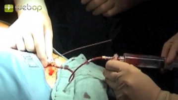
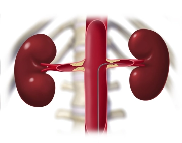
Advance a Terumo® guidewire through the sheath and introduce a pigtail catheter into the suprarenal aortic segment. Perform survey angiography of the aorta and renal artery segment in road-mapping technique. Angiography reveals bilateral subtotal stenosis of the renal arteries close to their origins. Next, in this mask place a Renal Double Curve (RDC) catheter into the suprarenal aortic segment. Slowly retract the guidewire until it slips into the infundibulum at the origin of the left renal artery. Once the guidewire is seated securely, exchange the Terumo® guidewire for a soft-tipped thinner, but harder, metal wire, advancing it carefully into the peripheral renal artery.
Tips:
1. Do not advance the metal wire too far. The resistance must be felt; otherwise the kidney will be perforated, which can result in severe parenchymal haemorrhage.
2. Why does the Terumo® catheter have to be exchanged for a stiffer metal catheter at all? This is necessary to achieve sufficient pushability for the balloon-expandable stent that is deployed in the next surgical step. A soft Terumo® guidewire would dislodge and the stent could not be positioned precisely because it would lack central purchase.
Roadmapping ("pathfinder"):
This involves administering a small bolus of contrast agent to display the abdominal aorta as a roadmap. This image is saved as a mask. The subsequent images are then acquired without contrast medium and subtracted from the mask. In this way, for example, only a radiopaque catheter with its current position can be displayed. In the resulting subtraction images, the bright catheter is visible against the dark abdominal aorta, and any background irrelevant to this examination is omitted.