Start your free 3-day trial — no credit card required, full access included

Anatomy - Right retrograde iliofemoral TEA (ring stripper desobliteration) with profunda patch plasty

  1. Arterial Supply of the Pelvis

    Arterial Supply of the Pelvis
    Zum Vergrößern bitte anklicken
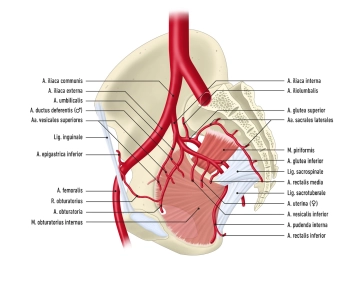
    • The abdominal aorta divides at the aortic bifurcation (approximately at the level of L4) into the two common iliac arteries
    • each common iliac artery in turn divides into an internal and external iliac artery
    • The internal iliac artery supplies mainly the pelvic viscera with visceral branches, and with its parietal branches it is involved in the supply of the lower extremities
    • The external iliac artery contributes to the supply of the pelvis and becomes the femoral artery after passing through the vascular lacuna
    1. Internal Iliac Artery

    Origin

    • arises immediately caudal to the aortic bifurcation from the common iliac artery

    Course

    • passes over the upper margin of the greater sciatic foramen caudally

    Positional Relationships

    • dorsal: internal iliac vein, piriformis muscle, lumbosacral plexus
    • ventral: ureter
    • lateral: external iliac vein, psoas major muscle
    • caudal: obturator nerve

    Branches

    visceral branches:

    • umbilical artery
    • inferior vesical artery
    • middle rectal artery
    • in women: uterine artery

    parietal branches:

    • iliolumbar artery
    • lateral sacral arteries
    • superior gluteal artery
    • inferior gluteal artery
    • obturator artery
    • internal pudendal artery

    Supply Area

    • pelvic organs, gluteal muscles
    1.1 Visceral Branches of the Internal Iliac Artery

    Main Branches

    Course and Branches

    Supply Area

    Umbilical Artery

    • postnatal obliteration to the medial umbilical ligament
    • remaining branches: superior vesical arteries, in men: artery of the ductus deferens
    • urinary bladder body
    • in men: ductus deferens

    Inferior Vesical Artery

    • branching in the bladder fundus into small branches
    • urinary bladder
    • in men: prostate, seminal vesicles

    Middle Rectal Artery

    • branching in the middle rectal section
    • anastomoses with superior and inferior rectal arteries
    • rectal ampulla
    • in women: vagina
    • in men: prostate

    Uterine Artery (Women)

    • passes over the ureter
    • tortuous course in the broad ligament of the uterus
    • gives off numerous small branches
    • ovary
    • uterine tube
    • proximal vagina
    • uterus
    1.2 Parietal Branches of the Internal Iliac Artery

    Main Branches

    Course and Branches

    Supply Area

    Iliolumbar Artery

    • runs obliquely upward to the iliopsoas muscle
    • psoas major muscle
    • iliacus muscle

    Lateral Sacral Arteries

    • superior branch: runs medially through the 1st or 2nd anterior sacral foramen and exits again through the posterior sacral foramen
    • inferior branch: passes over the piriformis muscle and medial to the sacral foramina on the sacrum caudally
    • sacral canal
    • piriformis muscle
    • muscles/skin dorsal to the sacrum

    Superior Gluteal Artery

    • passes with the superior gluteal nerve through the suprapiriform foramen and with branches to the gluteus maximus muscle (superficial branch) and gluteus medius muscle (deep branch)
    • gluteus maximus, medius, and minimus muscles
    • piriformis muscle
    • tensor fasciae latae muscle
    • skin over the sacrum

    Inferior Gluteal Artery

    • passes on the piriformis muscle caudally through the infrapiriform foramen
    • then runs together with the posterior femoral cutaneous nerve and sciatic nerve through the gluteus maximus muscle
    • gluteus maximus, medius, and minimus muscles
    • piriformis muscle
    • quadriceps femoris muscle
    • skin of the lower buttock and proximal thigh

    Obturator Artery

    • runs along the lateral pelvic wall forward and downward
    • branches to the ilium and iliacus muscle
    • after passing through the obturator canal, gives off further branches, including pubic branch (anastomoses with the inferior epigastric artery to form the corona mortis) and posterior branch (branch: acetabular artery -> artery of the femoral head)
    • ilium
    • iliacus muscle
    • thigh adductors
    • pubis
    • external hip muscles
    • femoral head

    Internal Pudendal Artery

    • runs ventral to the sacral plexus to the infrapiriform foramen
    • re-enters the lesser pelvis via the lesser sciatic foramen, through the pudendal canal, and branches in the ischiorectal fossa
    • distal third of the rectum
    • anal canal
    • urethra
    • scrotal skin/labia
    • bulb of the penis/vestibule
    • dorsum of the penis/clitoral prepuce
    2. External Iliac Artery

    Origin

    • arises caudal to the internal iliac artery from the common iliac artery

    Course

    • runs along the psoas major muscle to the inguinal ligament, through the vascular lacuna, and here becomes the femoral artery

    Positional Relationships

    • the right external iliac artery runs under the ureter

    Branches

    • deep circumflex iliac artery runs to the anterior superior iliac spine and anastomoses with the iliolumbar artery and superior gluteal artery
    • inferior epigastric artery runs medial to the deep inguinal ring cranially and between the transversus abdominis muscle and rectus abdominis muscle, further branches including cremasteric artery or artery of the round ligament of the uterus 

    Supply Area

    • ilium, lower portion of the abdominal wall
  2. Arterial Supply of the Lower Extremity

    1. Femoral artery

    A 320-2
    Zum Vergrößern bitte klicken
    1.1. Overview

    Origin

    • External iliac artery, which becomes the femoral artery after passing under the inguinal ligament

    Course

    • runs between iliopsoas muscle and pectineus muscle (= iliopectineal fossa) covered by fascia lata
    • is accompanied by the sartorius muscle up to the middle of the thigh
    • passes together with femoral vein and saphenous nerve through the adductor canal
    • transitions into the popliteal artery after passing through the adductor hiatus

    Branches

    • Superficial epigastric artery
    • Superficial circumflex iliac artery
    • Deep femoral artery (main artery for supplying the thigh!)
    • External pudendal arteries
    • Descending genicular artery

    Supply area

    • Thigh 
    • Skin of the abdominal wall
    • External genitalia
    • Knee joint and proximal/medial lower leg
    1.2. Important branches of the femoral artery

    Branches

    Supply area

    Superficial epigastric artery

    • Ø
    • Skin of the abdominal wall up to the umbilical region

    Superficial circumflex iliac artery

    • Ø
    • Anterior superior iliac spine

    Deep femoral artery

    • Medial circumflex femoral artery runs between iliopsoas muscle and pectineus muscle dorsally → trochanteric fossa; anastomosis with  lateral circumflex femoral artery
    • Lateral circumflex femoral artery runs between rectus femoris muscle and vasti muscles laterally; anastomosis with medial circumflex femoral artery
    • Perforating arteries pass through the adductors to the back of the thigh
    • Thigh

    External pudendal arteries

    • Ø
    • External genitalia
    • Scrotum or labia majora

    Descending genicular artery

    • Articular branches → genicular arterial network
    • Saphenous branch → runs medially together with great saphenous vein and saphenous nerve to the calf
    • Knee
    • Medial calf proximal

    2. Popliteal artery

    A 320-3
    Zum Vergrößern bitte anklicken
    2.1. Overview

    Origin

    • Continuation of the femoral artery after its passage through the adductor hiatus

    Course

    • runs through popliteal fossa
    • runs between the heads of the gastrocnemius muscle
    • transitions into the tibiofibular trunk after giving off the anterior tibial artery

    Branches

    • Anterior tibial artery

    Supply area

    • Popliteal fossa
    • Knee joint
    2.2. Important branches of the popliteal artery

    Course

    Branches

    Supply area

    Anterior tibial artery

    • Branch of the popliteal artery
    • is accompanied by 2 veins and in the lower part by the deep peroneal nerve
    • passes through the interosseous membrane of the leg and runs on its anterior side
    • runs behind the tendon of the extensor hallucis longus muscle
    • transitions into the dorsalis pedis artery
    • Posterior and anterior tibial recurrent arteries
    • Lateral and medial anterior malleolar arteries
    • Dorsalis pedis artery (terminal branch of the anterior tibial artery)
    • Anterior lower leg
    • Dorsum of the foot

    3. Posterior tibial artery

    3.1. Overview

    Origin

    • Continuation of the popliteal artery after the departure of the anterior tibial artery

    Course

    • runs together with the tibial nerve under the tendinous arch of the soleus muscle → medial malleolar groove → medial malleolus
    • Division into medial and lateral plantar arteries

    Branches

    • Fibular artery
    • Medial and lateral plantar arteries

    Supply area

    • Dorsal part of the lower leg
    • Sole of the foot
    3.2. Important branches of the posterior tibial artery

    Course

    Branches

    Supply area

    Fibular artery

    • runs on the back of the fibula 
    • runs between posterior tibial muscle and flexor hallucis longus muscle
    • Communicating branch: Transverse connection to the posterior tibial artery
    • Lateral malleolar branches
    • Lateral-dorsal lower leg

    Medial plantar artery

    • runs in the medial vascular-nerve bundle of the sole of the foot (medial plantar artery, vein, and nerve) between abductor hallucis muscle and flexor digitorum brevis muscle
    • Ø
    • Medial sole of the foot

    Lateral plantar artery

    • runs in the lateral vascular-nerve bundle of the sole of the foot (lateral plantar artery, vein, and nerve) between flexor digitorum brevis muscle and quadratus plantae muscle
    • Deep plantar arch: Anastomosis between lateral plantar artery and the deep plantar branch of the dorsalis pedis artery in the area of the metatarsal bones
    • Lateral sole of the foot
    • Toes