Start your free 3-day trial — no credit card required, full access included

Perioperative management - Right retrograde iliofemoral TEA (ring stripper desobliteration) with profunda patch plasty

  1. Indications

    According to the TASC criteria, therapeutic treatment options can be derived depending on the local occlusion/stenosis length. The length of the stenosis and its localization regions decide on the therapy: endovascular or open vascular surgery.

    TASC criteria aortoiliac vascular occlusions

    Types

    Morphology

    Therapy principle

    A

    Focal stenoses of the A. iliaca communis or A. iliaca externa  <3  cm, uni- or bilateral  

    endovascular

    B

    Focal stenoses 3–10 cm long and/or unilateral occlusion of the A. iliaca communis

    endovascular

    C

    Bilateral stenoses of the A. iliaca communis, 5–10 cm or unilateral complete occlusion of the A. iliaca externa or bilateral occlusions of the A. iliaca communis 

    open reconstruction

    D

    Diffuse stenotic changes of the entire iliac axis or unilateral occlusion of the A. iliaca communis and externa or bilateral occlusions of the A. iliaca externa  

    open reconstruction

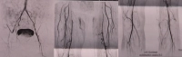
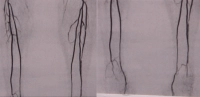
    Video example: PAD stage IIb right leg with subtotal stenosis of the external iliac artery, common femoral artery occlusion, profunda origin occlusion, long-segment occlusion of the superficial femoral artery, occlusion of the anterior tibial artery on both sides (right proximal, left peripheral) -> TASC D

    PM 320-1
    PM 320-2
    Zum Vergrößern bitte anklicken


    Fontaine stage

    Clinical presentation                                    

    Rutherford category

    Grade

    Clinical presentation

    I

    asymptomatic

    0

    0

    asymptomatic

    IIa

    Walking distance > 200 m

    1

    I

    mild claudication

    IIb

    Walking distance < 200 m

    2

    I

    moderate claudication


    3

    I

    severe claudication

    III

    ischemic rest pain

    4

    II

    ischemic rest pain

    IV

    ulcer, gangrene

    5

    III

    minor tissue loss

    6

    III

    major tissue loss

  2. Contraindications

    • ASA IV
    • severe COPD
    • Previous radiation in the groin
    • Infections in the area of the reconstruction      
  3. Preoperative Diagnostics

    History

    • Claudication
    • Walking distance
    • Risk factors -> Nicotine, arterial hypertension,  CHD, heart failure, diabetes mellitus, hyperlipidemia, manifest renal insufficiency with/without need for dialysis, coagulopathies

    Inspection

    • Skin changes
    • Muscular abnormalities
    • Orthopedic malpositions
    • Skin color
    • Hair growth
    • Trophic changes
    • Swelling, edema, mycoses, phlegmons, leg ulcers etc.    

    Side-comparing palpation

    • Pulse status
    • Skin temperature

    Side-comparing auscultation of the extremity arteries

    Palpation-Auskultation
    Palpation–auscultation

    Ankle-Brachial Index (ABI)

    • ABI = systolic BP posterior tibial artery/systolic BP brachial artery

    ABI value

    Severity of PAD

    > 1.3

    Falsely high values (suspicion of Mönckeberg mediasclerosis, e.g., in diabetes mellitus)

    > 0.9

    Normal finding

    0.75 - 0.9

    Mild PAD

    0.5 - 0.75

    Moderate PAD

    < 0.5

    Severe PAD

    • An ABI value of < 0.9 is considered proof of the presence of relevant PAD.
    • The determination of the ankle-brachial index (ABI) by non-invasive measurement of the Doppler occlusion pressure is a suitable test for detecting PAD.
    • For the diagnosis of PAD, the ABI value with the lowest ankle artery pressure is decisive.
    • A pathological ankle-brachial index is an independent risk indicator for increased cardiovascular morbidity and mortality.

    Color-coded duplex sonography

    • Carotid, abdominal aorta, extremity arteries
    • Localization of stenoses and occlusions in almost all vascular regions except in the thoracic area
    • Quantification of the degree of stenosis and assessment of plaque morphology possible
    • Sensitivity and specificity approx. 90%   
    • Well suited as a screening method

    Contrast-enhanced MR angiography or alternatively CT angiography

    • Validation of findings or for therapy planning

    Cardiac check

    • Resting ECG
    • Exercise ECG
    • Echocardiography

    Chest X-ray

    If necessary, spirometry

    Laboratory

    • CBC
    • Electrolytes
    • Coagulation
    • Retention values
    • Liver enzymes
    • Blood lipids
    • Blood group
  4. Special Preparation

    • Fast for 6 hrs. before the procedure
    • Mark the surgical side on the patient
    • Shave groin, lower abdomen and leg
    • Pack foot in cotton boot (Avoid intraoperative pressure damage when clamping the arteries)
    • AVK Stage IV: antibiotic treatment according to antibiogram and continuation of therapy postoperatively for at least 5 days (depending on local findings)
  5. Informed Consent

    General Surgical Risks

    • Severe bleeding, blood transfusions, transmission of Hepatitis/HIV through blood products
    • Allergy/Intolerance
    • Wound infection
    • Thrombosis/Embolism
    • Skin, vascular, nerve damage e.g. due to positioning
    • Keloids

    Specific Surgical Risks

    • renewed vascular occlusion, possibly further intervention, (partial) amputation
    • persistent ischemia with risk of amputation
    • Vascular dissection/rupture due to balloon catheter, possibly stent or interposition from vein or synthetic material
    • massive infections with severe bleeding from suture sites, sepsis, amputation
    • Nerve injury with paresthesia or pain, weakness or partial paralysis of the extremity
    • Embolism when withdrawing the balloon catheter, e.g. gangrene in the foot area, amputation
    • Lymphedema, lymphocele, -fistula
    • Compartment syndrome
    • Restriction of kidney function due to contrast medium in the context of intraoperative angiography
    • Risk of ureteral lesion if retroperitoneal preparation is necessary
    • in case of failure of TEA, method change with prosthetic replacement (uni- or also bilateral e.g. Y-prosthesis)
  6. Anesthesia

    • Intubation anesthesia
    • Spinal anesthesia (Caution: a newly occurring ischemia in postoperative occlusion of the reconstruction may possibly not be noticed by the patient!)
  7. Positioning

    Positioning
    • Supine position
    • right arm tucked, left arm extended
  8. OR Setup

    OR Setup

    The surgeon stands on the side to be operated on (in this case, on the right), opposite him the assistant. To the left of the assistant, the OR specialist is positioned.  The mobile DSA unit  is brought in from the left side if needed, the monitor is located in a clearly visible position for the surgeon at the patient's head height.

  9. Special Instruments and Holding Systems

    • various wound retractors
    • 120° angle clamps
    • Profunda clamp
    • Bulldog clamps
    • Potts scissors
    • button cannulas of different sizes
    • ring stripper, Fogarty catheter (various sizes or calibers)
    • 6F sheath (for intraoperative angiography)
    • monofilament suture material 5-0 and 6-0 with atraumatic needle
    • Dacron patch
    • vessel loops
    • moist abdominal cloths for protection of the wound edges
    • vascular prostheses of different designs, various calibers
    • instrumentation for intervention if necessary (dilation catheters, guide wires, stents)
    • mobile DSA unit
  10. Postoperative Treatment

    Postoperative Analgesia

    Follow the link here to PROSPECT (Procedure Specific Postoperative Pain Management) and to the current guideline Treatment of acute perioperative and posttraumatic pain.

    Medical Follow-up Care

    • 24-hour follow-up care in intensive care unit, possibly Intermediate Care
    • Normal ward possible from 1st-3rd day postoperatively
    • Close monitoring of heart-circulation-lungs
    • Check pulses in the legs, capillary perfusion of the feet
    • Therapeutic heparinization weight-adapted; on the day of surgery only 10,000 IU from the 4th postoperative hour (risk of rebleeding), then increase dose; after 4-5 days overlapping antiplatelet agents; possibly modification depending on cardiac risk profile (consult with cardiology)
    • Mobilization
    • from the 2nd-3rd day with help to the edge of the bed
    • Walking exercises only after 5 days
    • Caution with limited mobility/gait insecurity: In case of falls, the arterial patch can tear!

    Physiotherapy

    • isometric exercises
    • careful mobilization see above

    Diet Buildup

    • after 4-6 hours

    Stool Regulation

    • usually not required

    Incapacity for Work

    • approx. 6 weeks