Start your free 3-day trial — no credit card required, full access included

Anatomy - Transfemoral Fogarty balloon catheter embolectomy in embolic occlusion of the left external iliac artery—Vascular Surgery

  1. Arterial pelvic blood supply

    Arterial pelvic blood supply
    Zum Vergrößern bitte klicken
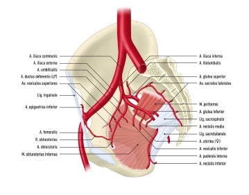
    • The abdominal aorta divides at the aortic bifurcation (around level L4) into both common iliac arteries
    • In turn, each common iliac artery divides into an internal and external iliac artery
    • With its visceral branches, the internal iliac artery supplies mainly the pelvic organs, while its parietal branches ensure the blood supply to the lower extremities
    • The external iliac artery contributes to the pelvic blood supply and, after passing through the vascular compartment (lacuna vasorum retroinguinalis), becomes the femoral artery
    1. Internal iliac artery

    Origin

    • Arises from the common iliac artery immediately inferior to the aortic bifurcation

    Course

    • Courses caudad over the superior border of the greater sciatic foramen

    Relation

    • Posterior: internal iliac vein, pirirformis muscle, lumbosacral plexus
    • Anterior: ureter
    • Lateral: external iliac vein, psoas major
    • Inferior: obturator nerve

    Branches

    Visceral branches:

    • Umbilical arteries
    • Inferior vesical artery
    • Middle rectal artery
    • In women: Uterine artery

    Parietal branches:

    • Iliolumbar artery
    • Lateral sacral arteries
    • Superior gluteal artery
    • Inferior gluteal artery
    • Obturator artery
    • Internal pudendal artery

    Distribution

    • Pelvic organs, gluteal muscles
    2. External iliac artery

    Origin

    • Arises from the common iliac artery immediately distal to the origin of the internal iliac artery

    Course

    • Courses along the major psoas muscle to the inguinal ligament, and after passing through the vascular compartment becomes the femoral artery

    Relation

    • The right external iliac artery courses posterior to the ureter

    Branches

    • The deep iliac circumflex artery courses to the superior iliac spine and joins the Iliolumbar artery and superior gluteal artery
    • The inferior epigastric artery runs craniad medial to the deep inguinal ring and between the transverse abdominis and rectus abdominis muscles; other branches include the cremasteric artery and artery of round ligament of uterus 

    Distribution

    • Iliac bone, inferior aspect of abdominal wall
  2. Arterial blood supply to the lower extremity

    1. Femoral artery
    A 307-2
    Zum Vergrößern bitte klicken
    1,1. Overview

    Origin

    • External iliac artery which becomes the femoral artery after passing posterior to the inguinal ligament

    Course

    • Passes between the iliopsoas and pectineus muscles (= iliopectineal fossa) covered by fascia lata
    • Accompanied as far as the mid-thigh by the sartorius muscle
    • Passes together with the femoral vein and saphenous nerve through the adductor canal (Hunter)
    • Becomes the popliteal artery after passing through the adductor hiatus

    Branches

    • Superficial epigastric artery
    • Superficial iliac circumflex artery
    • Deep femoral artery (main artery supplying the thigh!)
    • External pudendal arteries
    • Descending genicular artery

    Distribution

    • Thigh 
    • Abdominal wall skin
    • External genitalia
    • Knee joint and proximal/medial lower leg
    1,2. Major branches of femoral artery

    Branches

    Distribution

    Superficial epigastric artery

    • Ø
    • Abdominal wall skin up to the umbilical region

    Superficial iliac circumflex artery

    • Ø
    • Superior anterior iliac spine

    Deep femoral artery

    • The medial femoral circumflex artery courses posteriad between iliopsoas and pectineus muscles → trochanteric fossa; anastomosis with lateral femoral circumflex artery
    • The lateral femoral circumflex artery runs laterad between the rectus femoris and vastus muscles; anastomosis with the medial femoral circumflex artery
    • The perforating arteries course through the adductors to the posterior aspect of the thigh
    • Thigh

    External pudendal arteries

    • Ø
    • External genitalia
    • Scrotum / major pudendal labia

    Descending genicular artery

    • Articular branches → Rete articulare genus
    • Saphenous branch → courses medially to calf together with the great saphenous vein and saphenous nerve
    • Knee
    • Proximal medial calf

    2. Popliteal artery

    A 307-3
    Zum Vergrößern bitte klicken
    2,1. Overview

    Origin

    • Continuation of femoral artery after it passes through the adductor hiatus

    Course

    • Runs through popliteal fossa
    • Passes between gastrocnemius heads
    • Becomes the tibiofibular trunk, after giving rise to the anterior tibial artery

    Branches

    • Anterior tibial artery

    Distribution

    • Popliteal fossa
    • From top of the patella to center of the knee joint
    2,2. Major branches of popliteal artery

    Course

    Branches

    Distribution

    Anterior tibial artery

    • Branch of popliteal artery
    • Accompanied by two veins and, in its inferior aspect, by deep peroneal nerve
    • Passes through interosseous membrane of leg and courses on its anterior aspect
    • Passes posterior to extensor hallucis longus tendon
    • Becomes the dorsalis pedis artery
    • Posterior + anterior tibial recurrent artery
    • Lateral + medial anterior malleolar artery
    • Dorsalis pedis artery (terminal branch of anterior tibial artery)
    • Anterior lower leg
    • Dorsum of foot
    3. Posterior tibial artery
    3,1. Overview

    Origin

    • Continuation of popliteal artery distal to origin of anterior tibial artery

    Course

    • Passes together with tibial nerve posterior to tendinous arch of soleus → Medial malleolar sulcus → Medial malleolus
    • Bifurcates into medial + lateral plantar artery

    Branches

    • Fibular artery
    • Medial + lateral plantar artery

    Distribution

    • Dorsal aspect of lower leg
    • Sole of foot
    3,2. Major branches of posterior tibial artery

    Course

    Branches

    Distribution

    Fibular artery

    • Courses along dorsal aspect of fibula 
    • Lies posterior tibial and flexor hallucis longus muscles
    • Communicating branch: Cross-connection to posterior tibial artery
    • Lateral malleolar branches
    • Lateroposterior lower leg

    Medial plantar artery

    • Courses in medial neurovascular bundle of sole of foot (medial plantar artery, vein and nerve) between abductor hallucis and flexor digitorum brevis muscles
    • Ø
    • Medial sole of foot

    Lateral plantar artery

    • Courses in lateral neurovascular bundle of sole of foot (lateral plantar artery, vein and nerve) between flexor digitorum brevis and quadratus plantae muscles
    • Deep plantar arch: anastomosis between lateral plantar artery and deep plantar branch of dorsalis pedis artery at the metatarsals
    • Lateral sole of foot
    • Toes