Start your free 3-day trial — no credit card required, full access included

Perioperative management - Transfemoral Fogarty balloon catheter embolectomy in embolic occlusion of the left external iliac artery—Vascular Surgery

  1. Indications

    • Arterial embolism resulting in acute limb ischemia

    80-90% of all peripheral emboli originate from thrombi in the left atrium, and 70% of all peripheral emboli involve the aortoiliac axis. Vascular branches are most often affected, especially the femoral bifurcation and the popliteal level.

    More than 70% of cardiac embolisms are by absolute arrhythmia in atrial fibrillation. Other sources of cardiac embolisms:  acute myocardial infarction (5%); dilated cardiomyopathy; valve defects; endocarditis; prosthetic heart valve replacement; heart wall aneurysm; and atrial myxoma.

    10-20% of all peripheral thromboembolism are caused by non-cardiac sources of embolism: Aneurysms of the aortoiliac and femoropopliteal axis (microembolism -> "blue toe syndrome" or "trash foot" ); arteriosclerotic plaques; rarely tumors (lung cancer, pulmonary metastases with connection to the pulmonary circulation, angiosarcoma); foreign bodies; or paradoxical embolism in patent foramen ovale cordis.

    In 5-10% of cases, the origin of a peripheral arterial embolism is unknown.

  2. Contraindications

    • ASA IV
  3. Preoperative diagnostic work-up

    The diagnosis of acute limb ischemia can often be made "at a glance" after a brief history and examination. Normally, angiography is not performed (exception: concurrent PAOD) and management is planned based on the clinical findings (history, inspection, palpation). After having resolved the absolute ischemia, diagnosis and treatment of the source of the embolism can be approached in orderly fashion.

    Emergent diagnostic work-up should address the following questions:

    1. Severity of ischemia (Pratt's 6 Ps, TASC in PAOD)?
    2. Localization and extent of the occlusion?
    3. Acute ischemia resulting from embolization or PAOD?

    The typical 6 Ps of Pratt characterize the severe course of acute limb ischemia with grave threat to the limb viability (Source: Pratt GH (1954) Cardiovascular surgery. Kimpton, London):

    Pratt‘s six Ps

    Significance

    Pulselesness

    lLck of peripheral pulses

    Pallor

    Paleness of the skin

    Pain

    Increasing ischemic pain

    Paresthesia

    Ascending sensory deficit

    Paralysis

    Increasing functional impairment

    Prostration

    Progressive tissue destruction

    In PAOD, the classification of acute limb ischemia according to the Transatlantic Inter-Society Consensus (TASC) Working Group seeks to reflect the varying levels of urgency for further diagnostic work-up and management:

    Stage

    Description

    TASC I

    • No threat to limb viability
    • Sensory and motor function preserved
    • Peripheral pulses detectable by Doppler ultrasonography

    TASC IIa

    • Mild impairment of motor and sensory function
    • Peripheral pulses usually detectable by Doppler ultrasonography

    TASC IIb

    • Substantial threat to limb viability
    • Loss of sensory function
    • Pain at rest proximal to the toes
    • Peripheral pulses usually not detectable by Doppler ultrasonography

    TASC III

    • Irreversible tissue destruction or severe peripheral nerve damage
    • Severe sensory impairment
    • Limb paralysis

    Medical history/clinical picture

    Rough distinction between acute thrombotic occlusion and embolism:

     Embolism

    • Sudden, occasionally whip-like pain in the limb (cardinal symptom)
    • Often severe ischemia
    • Cardiovascular history: known atrial fibrillation; heart defect; history of myocardial infarction; aortic aneurysm

    → thrombotic occlusion

    • more likely symptoms increasing over several days, typically with incomplete ischemia
    • Known PAOD (claudication symptoms?)
    • History of bypass surgery, stenting/PTA
    • Atherosclerosis risk profile
    • Local trauma
    • No evidence of source of the embolism

    Skin color

    • White ischemia: pale distal to the occlusion
    • Blue ischemia: less favorable prognosis because thrombus formation due to stasis has already spread to the venous circulation
    • Spontaneous Ratschow test may be seminal in incomplete ischemia (total pallor with the limb elevated and no recovery even after returning the limb to the horizontal position).

    Capillary refill time

    • short plantar pressure with finger on big toe or forefoot → initially pale pressure point usually turns red within < 3 seconds
    • The longer the redness takes to appear, the more marked is the perfusion disorder

    Effective perfusion pressure

    • Elevation of the limb allows the perfusion pressure to be estimated → 10 cm = 7.5 mmHg

    Skin temperature

    • The affected limb is colder when comparing both extremities
    • The occlusion is much more proximal than the drop in temperature
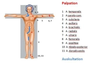
    Pulses

    • In the simplest case, lack of pulses in the affected limb
    • Bilateral lack of pulses: pre-existing bilateral PAOD or asymptomatic embolism

    Auscultatory bilateral comparison of the limb arteries

    PM 303-3

    Ankle-Brachial Index (ABI)

    • ABI = systolic BP of posterior tibial artery / systolic BP of brachial artery
    • the smaller the ABI, the more pronounced the ischemia
    • In acute stages, the pressure might not be measurable

    Color-flow Doppler ultrasonography

    • Carotid artery, abdominal aorta, limb arteries (rule out popliteal aneurysm in lower leg occlusion!)
    • Stenoses and occlusions in almost all vascular regions apart from chest
    • Allows quantifying the degree of stenosis and assessing plaque morphology
    • Sensitivity and specificity around 90%   

    CT angiography

    • Multislice computed tomography (MS-CT) with nonionic contrast agent
    • Broad range of indications: traumatic vascular lesion (esp. trunk); vascular dissection/rupture; aneurysm; arterial thrombosis/embolism; portal vein/mesenteric vein thrombosis; pulmonary artery embolism; PAD; vascular tumors
    • Benefits: rapid; detects relevant comorbidities; visualizes peripheral arteries; sensitivity and specificity each about 90%
    • Drawbacks: Radiation and contrast agent exposure, allergies (about 3%), no functional assessment
    • Indication does not depend on the degree of ischemia but on the history and clinical findings in the contralateral leg: angiography is recommended in case of pre-existing PAOD or evidence of popliteal aneurysm. The findings then determine the surgical strategy. The presence of concomitant PAOD or popliteal aneurysm as the origin of acute ischemia necessitates more complex repair (e.g., interposition grafts, bypass procedures). However, in case of complete ischemia do not waste too much time on angiography (suitable logistics).

    Laboratory panels

    • Blood count
    • Electrolytes
    • Coagulation
    • Kidney function parameters
    • Liver function parameters
    • Blood lipids
    • Blood group

    Cardiac check

    • Resting ECG

    Chest x-ray

    As an emergency measure administer 5000-10000 IU heparin to prevent appositional thrombosis. Protect the foot in a padded bootie to prevent pressure injury (especially during surgery as well!).

  4. Special preparation

    Acute limb ischemia is a vascular emergency, so measures taken as part of special preparation should be limited to the bare essentials.

    • Shaving the surgical field
    • While packed RBCs are usually not required, the blood group should be known.
    • In advanced ischemia and acute threat to limb viability and patient life, the fasting criteria may not be met
  5. Informed consent

    General surgical risks

    • Major bleeding, blood transfusions, transmission of hepatitis/HIV through allogeneic blood transfusions
    • Allergy/intolerance
    • Wound infection
    • Thrombosis/embolism
    • Skin/vascular/nerve damage, e.g., due to patient positioning
    • Keloid

    Procedure-specific risks

    • Repeated vascular occlusion, possibly repeat procedure, (partial) amputation
    • Vascular dissection/rupture by the balloon catheter, mange by stenting or venous interposition or prosthetic graft, if necessary
    • Massive infections with severe bleeding from the suture lines, sepsis, amputation
    • Nerve injury with paresthesia or pain, weakness or partial limb paralysis
    • Embolism during withdrawal of the balloon catheter, e.g., gangrene in the foot, amputation
    • Lymphedema
    • Impaired renal function induced by contrast agent during intraoperative angiography
  6. Anesthesia

    Local anesthesia with an anesthesiologist on “stand-by” or General anesthesia

  7. Positioning

    Positioning

    Supine, atm tucked in on the affected side, contralateral arm abducted

  8. Operating room setup

    Operating room setup

    Surgeon on the affected side, with the assistant and scrub nurse opposite

  9. Special instruments and fixation systems

    • Standard vascular instrument tray
    • 5F Fogarty arterial embolectomy catheter (white): External and common iliac artery
    • 4F Fogarty arterial embolectomy catheter (red): Cubital and brachial artery, common/deep femoral artery (proximal), superficial femoral artery and popliteal artery
  10. Postoperative management

    Postoperative analgesia:

    Follow this link to PROSPECT (Procedures Specific Postoperative Pain Management) and to the current German guideline Behandlung akuter perioperativer and posttraumatischer Schmerzen [Treatment of acute perioperative and posttraumatic pain].

    Postoperative care

    • 24-hour monitoring on ICU or possibly intermediate care unit
    • Regular ward possible from postoperative day 1 to 3
    • Close cardiovascular and pulmonary monitoring
    • Monitoring of limb pulses, duplex ultrasonography
    • Weight-adapted therapeutic heparinization; on day of surgery, only 10,000 IU starting on the 4th postoperative hour 4 (risk of secondary bleeding), then increase heparin dose to avoid reembolization until source of embolism has been identified
    • Coumarins or dabigatran only once wound healing is complete 

    Ambulation

    • Starting on postoperative day 1 sitting on edge of bed
    • Starting on postoperative 3 cautious assisted getting out of bed

    Physical therapy

    • Isometric exercises
    • Breathing exercises

    Diet

    • 4-6 hrs. postop., depending on patient condition

    Bowel movement

    • Assist starting postoperative day 3, if needed

    Work disability

    • Case by case, depending on the clinical picture

    Do not forget the diagnostic work-up and managment of the source of the embolism!