
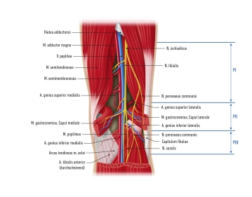
The popliteal artery (PA) is 12–18 cm long and has a diameter of about 9 mm, which tapers down to about 5 mm towards the periphery. As the direct continuation of the superficial femoral artery, the PA originates above the knee (suprageniculate) in the adductor hiatus, where it is enclosed laterally by the biceps femoris and medially by the semimembranosus muscles (femoral space). In the popliteal fossa, the PA lies between the femoral condyles in the gastrocnemius tunnel, where it is protected medially by the muscle and tendon complex of the pes anserinus. The infrageniculate PA courses into the so-called tibial space, crosses the popliteus muscle and divides at its lower border.
Important collateral branches (rete articulare genus) originate in the middle segment of the PA:
- Sural arteries
- Middle genicular artery
- Medial superior and inferior genicular arteries
- Lateral superior and inferior genicular arteries
Approximately 7 cm below the joint space of the knee at the level of the tendinous arch of the soleus muscle (arcus tendineus musculi solei), the PA divides into the anterior tibial artery and the tibiofibular trunk, marking the beginning of the crural artery segment.
Anatomical variants are present in 5%–8% of cases. These are mainly variant origins of the tibial arteries and positional variants of the PA in relation to the gastrocnemius or popliteus muscle.
In terms of surgical technique, the PA is usually divided into three segments (PI–PIII), which determine the surgical approaches:
Segment | Feature | Access |
|---|---|---|
PI | From the end of the adductor canal to the gastrocnemius tunnel proximal to the knee joint | Distal medial thigh |
PII | Knee joint | Posterior (popliteal fossa) |
PIII | From the infrapopliteal end of the gastrocnemius tunnel to the origin of the anterior tibial artery/tendinous arch of the soleus muscle | Proximal medial lower leg |
The PA courses in a neurovascular bundle surrounded by fatty connective tissue (corpus adiposum popliteum). In the PI and PII segments the artery lies anteromedial to the popliteal vein (PV), whereas in the PIII segment it is surrounded by the venae comitantes. The tibial nerve and common peroneal nerve lie posterolateral to the PA and PV. In the popliteal fossa, the nerves have a relatively superficial course, which must be taken into account when exposing the PA from the posterior aspect.
The superficial and deep lymphatic drainage in the popliteal fossa is bundled and drained via three to five perivenously located lymph nodes.
Owing to its location, the PA is exposed to considerable mechanical stress, which is why, as a muscular-type artery, it possesses strong muscular and fibrous layers (tunica media, tunica elastica interna). The properties of its walls therefore resemble those of central elastic arteries.