Start your free 3-day trial — no credit card required, full access included

Anatomy - TEA of the femoral bifurcation, profunda patch plasty, dilatation and stent placement of the left superficial femoral artery

  1. Femoral artery

    Femoral artery
    Zum Vergrößern bitte klicken
    1.1. Overview

    Origin

    • External iliac artery, which becomes the femoral artery after passing under the inguinal ligament

    Course

    • runs between M. iliopsoas and M. pectineus (= iliopectineal fossa) covered by fascia lata
    • is accompanied by the M. sartorius up to the middle of the thigh
    • passes together with V. femoralis and N. saphenus through the adductor canal
    • transitions into the popliteal artery after passing through the adductor hiatus

    Branches

    • Superficial epigastric artery
    • Superficial circumflex iliac artery
    • Deep femoral artery (main artery for supplying the thigh!)
    • External pudendal arteries
    • Descending genicular artery

    Supply area

    • Thigh 
    • Skin of the abdominal wall
    • external genitalia
    • Knee joint and proximal/medial lower leg
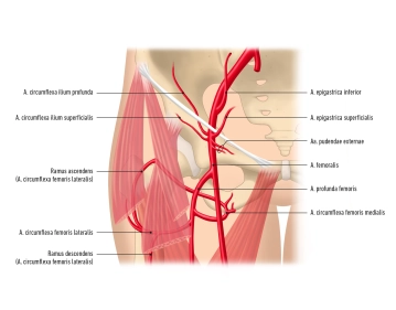
    1.2. Important branches of the femoral artery

    Branches

    Supply area

    Superficial epigastric artery

    • Ø
    • Skin of the abdominal wall up to the umbilical region

    Superficial circumflex iliac artery

    • Ø
    • Anterior superior iliac spine

    Deep femoral artery

    • Medial circumflex femoral artery runs between M. iliopsoas and M. pectineus dorsally → trochanteric fossa; anastomosis with  lateral circumflex femoral artery
    • Lateral circumflex femoral artery runs between M. rectus femoris and Mm. vasti laterally; anastomosis with medial circumflex femoral artery
    • Perforating arteries pass through the adductors to the back of the thigh
    • Thigh

    External pudendal arteries

    • Ø
    • external genitalia
    • Scrotum or labia majora

    Descending genicular artery

    • Articular branches → genicular articular network
    • Saphenous branch → runs medially together with great saphenous vein and saphenous nerve to the calf
    • Knee
    • proximal medial calf