Start your free 3-day trial — no credit card required, full access included

Perioperative management - TEA of the femoral bifurcation, profunda patch plasty, dilatation and stent placement of the left superficial femoral artery

  1. Indications

    The consensus document TASC II (Transatlantic Inter-Society Consensus for the Management of Peripheral Arterial Disease) deals with aspects of revascularization in PAD.

    According to the TASC criteria, therapeutic treatment options can be derived depending on the local occlusion/stenosis length. The length of the stenosis and its localization regions decide on the therapy: endovascular or open vascular surgery.

    TASC criteria for femoropopliteal vascular occlusions

    Types

    Morphology

    Therapy principle

    A

    Single stenosis  <5  cm length, not at the origin of the SFA or in distal popliteal artery, single occlusion  <3  cm length (not at the origin of the SFA or popliteal artery)  

    endovascular

    B

    Single stenosis 5–10 cm length, not in distal popliteal artery, single occlusion 3–10 cm length, not in distal popliteal artery, calcified stenosis  <5  cm length, multiple lesions  <3  cm length

    endovascular

    C

    Single occlusion 3–10 cm length up to distal popliteal artery, multiple focal lesions 3–5 cm length without/with calcification, single stenosis/occlusion  >10 cm length  

    open reconstruction

    D

    Complete occlusion of CFA and/or SFA, complete occlusion of popliteal artery and trifurcation, severe diffuse disease

    open reconstruction

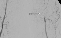
    Video example:  TASC C → open reconstruction

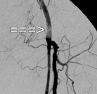
    PAD stage IIb left (walking distance under 100 m). The preoperative DSA shows:

    • a subtotal stenosis of the common femoral artery
    • a long-segment occlusion of the superficial femoral artery in the adductor canal
    • partial occlusions of the lower leg arteries
    PM 322-1
    Subtotale Stenose der Arteria femoralis communis
    PM 322-2
    Langstreckigen Verschluss der Arteria femoralis superficialis
    PM 322-3
    Teilverschlüsse der Unterschenkelarterien
  2. Contraindications

    • Infections in the area of the reconstruction
    • ASA IV
    • Status post radiation in the groin (then extra-anatomical bypass routing required)
  3. Preoperative Diagnostics

    History

    • Claudication
    • Walking distance
    • Risk factors → Smoking, arterial hypertension,  CHD, heart failure, diabetes mellitus, manifest renal insufficiency with/without need for dialysis, coagulopathies

    Inspection

    • Skin changes
    • Muscular abnormalities
    • Orthopedic malpositions
    • Skin color
    • Hair growth
    • Trophic changes
    • Swelling, edema, mycoses, phlegmons, leg ulcers etc.    

    Bilateral comparative palpation

    • Pulse status
    • Skin temperature

    Bilateral comparative auscultation of the extremity arteries

    Palpation-Auskultation
    Palpation–auscultation

    Ankle-Brachial Index (ABI)

    • ABI = systolic BP posterior tibial artery/systolic BP brachial artery

    ABI value

    Severity of PAD

    > 1.3

    Falsely high values (suspicion of Mönckeberg mediasclerosis, e.g., in diabetes mellitus)

    > 0.9

    Normal finding

    0.75 - 0.9

    Mild PAD

    0.5 - 0.75

    Moderate PAD

    < 0.5

    Severe PAD

    • An ABI value of < 0.9 is considered proof of the presence of relevant PAD.
    • The determination of the ankle-brachial index (ABI) by non-invasive measurement of the Doppler occlusion pressure is a suitable test for detecting PAD.
    • For the diagnosis of PAD, the ABI value with the lowest ankle artery pressure is decisive.
    • A pathological ankle-brachial index is an independent risk indicator for increased cardiovascular morbidity and mortality.

    Color-coded duplex sonography

    • Carotid, abdominal aorta, extremity arteries
    • Localization of stenoses and occlusions in almost all vascular regions except in the thoracic area
    • Quantification of the degree of stenosis and assessment of plaque morphology possible
    • Sensitivity and specificity approx. 90 %   
    • Well suited as a screening method

    MR angiography, if necessary i.a. DSA (in complex changes)

    Cardiac check

    • Resting ECG
    • Exercise ECG
    • Echocardiography

    Chest X-ray

    If necessary, spirometry

    Laboratory

    • CBC
    • Electrolytes
    • Coagulation
    • Renal function tests
    • Liver enzymes
    • Blood lipids
    • Blood type
    • In PAD stage IV → wound swab/antibiogram
  4. Special Preparation

    • Shortening of the hair in the surgical area
    • Order blood units
    • Pack the foot of the side to be operated in a cotton boot (avoidance of intraoperative pressure injuries during clamping of the vessels)
    • Antibiotic therapy according to antibiogram (see Diagnostics) for 5 days postoperatively (depending on local findings)
  5. Informed Consent

    General Surgical Risks

    • Severe bleeding, blood transfusions, transmission of hepatitis/HIV through donor blood products
    • Allergy/Intolerance
    • Wound infection
    • Thrombosis/Embolism
    • Skin, vascular, nerve damage e.g. due to positioning
    • Keloids

    Specific Procedure Risks

    • Vascular occlusion due to thrombosis; surgical revision, (partial) amputation
    • massive infections with severe bleeding from suture sites of the patch
    • Injury to sensory and motor nerves; dysesthesias, pain, temporary or also persistent (partial) paresis of the thigh muscles
    • Damage to lymphatic vessels;  temporary or persistent lymphedema, lymph fistula
    • Compartment syndrome; if necessary, surgical decompression/fasciotomy, persistent paresis, loss of extremity
    • Renal function impairment with temporary or permanent dialysis due to tourniquet syndrome and intraoperative angiography (contrast medium)
    • Suture site aneurysm; surgical intervention depending on size, clinical presentation
  6. Anesthesia

    • ITN
    • Spinal anesthesia (Caution: Pat. may not notice a newly occurring ischemia in case of postoperative early closure of the reconstruction!)
  7. Positioning

    Positioning
    • Supine position
    • contralateral arm abducted (in principle, abduction of both arms is also possible)
  8. OR Setup

    OR Setup

    The surgeon stands on the side to be operated on, opposite him the assistant. The OR specialist stands to the right next to the assistant. The C-arm is brought in from the opposite side – here right – if needed, the monitor is located at the foot end of the patient clearly visible to the surgeon.

  9. Special Instrumentation and Holding Systems

    Atraumatic vascular instrumentation with:

    • various wound retractors
    • 120° angle clamps, profunda clamp
    • Pott's scissors
    • button cannulas of different sizes
    • 7F sheath
    • moist abdominal cloths for protection of the wound edges
    • monofilament suture material 5-0 and 6-0, atraumatic
    • Dacron patch
    • Vessel loops
    • if necessary, instrumentation for intervention (dilation catheters of different sizes, guide wires, stents)
    • heparin saline solution
    • mobile DSA unit
  10. Postoperative Treatment

    Postoperative Analgesia

    Follow the link here to PROSPECT (Procedures Specific Postoperative Pain Management) and to the current guideline Treatment of acute perioperative and posttraumatic pain.

    Medical Follow-up Care

    • depending on risk profile, short stay in Intermediate Care, possibly immediately to normal ward
    • Check pulses in the legs, capillary perfusion of the feet

    Heparin

    • Heparin weight-adapted i.v., at least 10,000 IU over 24 hours
    • Start 4 hours postoperatively
    • after 4-5 days switch to platelet aggregation inhibitors
    • possibly modification in case of concomitant CHD (consult with cardiologist)

     Mobilization

    • from day 1 postoperatively with assistance to the edge of the bed
    • Walking exercises only after 2-3 days

     Physiotherapy

    • possibly respiratory exercises for COPD
    • isometric muscle training

    Diet Build-up

    • 4-6 hours postoperatively

    Bowel Regulation

    • usually unnecessary

    Inability to Work

    • approx. 6 weeks