Origin |
|
|---|---|
Course |
|
Division |
|
Vascular territory | 1. Internal carotid artery
2. External carotid artery
|
-
Common carotid artery
![Common carotid artery]()
Zum Vergrößern bitte anklicken -
Internal carotid artery
Origin
- Branch of the common carotid artery (carotid bifurcation)
Course from caudal to cranial
1. Cervical part
- Section between origin of the common carotid artery to the skull base
- entry into the cranial cavity via the carotid canal
- gives off no branches
2. Petrous part
- runs in the petrous bone
- small branches to the tympanic cavity and pterygoid canal
3. Cerebral part
- runs in the subarachnoid space
- passes through the dura mater
- Branching into anterior cerebral artery and middle cerebral artery
- Anterior cerebral artery communicates via the anterior communicating artery with the contralateral anterior cerebral artery
The circle of Willis is an arterial vascular ring at the base of the brain, which serves the blood supply of the brain. It is composed – from anterior to posterior - of the following vessels:
- Anterior communicating artery (unpaired)
- Anterior cerebral artery (left and right)
- Internal carotid artery (left and right) or its direct continuation, the middle cerebral artery
- Posterior communicating artery (left and right)
- Posterior cerebral artery (left and right, both from the basilar artery).
However, there are numerous variants in the formation of the circle regarding the caliber of the branches and the connections (hypoplasias of individual branches or even agenesis of partial sections). This is clinically relevant for collateral supply in stenoses.
-
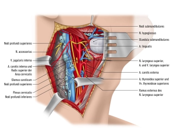
External carotid artery
The supply areas of the external carotid artery can be divided into four groups depending on location: anterior, middle and posterior group as well as terminal branches.
1. Anterior Group
Branches (cranial → caudal)
Branches
Supply
Superior thyroid artery
- Infrahyoid branch
- Cricothyroid branch
- Sternocleidomastoid branch
- corresponding muscles
- Superior laryngeal artery
- Inner side of larynx
- Glandular branches
- Thyroid gland
Lingual artery
- Suprahyoid branch
- Hyoid bone
- Dorsal lingual branches
- Dorsum of tongue
- Sublingual artery
- Sublingual gland
- Deep lingual artery
- Tip of tongue
Facial artery
- Ascending palatine artery
- soft palate
- tonsils
- pharynx
- Submental artery
- Submandibular gland
- suprahyoid muscles
- Inferior labial artery
- Lower lip
- Superior labial artery
- Upper lip
- Angular artery
- medial canthus
2. Middle Group
Branches (cranial → caudal)
Branches
Supply
Ascending pharyngeal artery
- Pharyngeal branches
- Pharynx
- Inferior tympanic artery
- Tympanic cavity
- Posterior meningeal artery
- Dura mater
3. Posterior Group
Branches (cranial → caudal)
Branches
Supply
Occipital artery
- Mastoid branch
- Mastoid cells
- Occipital branches
- Occipital region
- Meningeal branch
- Dura mater
Posterior auricular artery
- Auricular branch
- external ear
- Occipital branch
- Occipital region
- Stylomastoid artery
- Facial nerve
- Tympanic cavity
- Mastoid cells
- Posterior tympanic arteries
- Tympanic cavity
- Mastoid cells
- Pharyngeal branches
- Pharynx
- Parotid branch
- Parotid gland
4. Terminal branches
Branches (cranial → caudal)
Supply
Superficial temporal artery
- Transverse facial artery
- Face
- Zygomatico-orbital artery
- lateral canthus
- Middle temporal artery
- Temporal muscle
- Frontal branch
- Scalp
Maxillary artery
Mandibular part
- Deep auricular artery
- Temporomandibular joint
- external auditory canal
- Anterior tympanic artery
- Tympanic cavity
- Inferior alveolar artery
- Teeth
- Mandible
- Mylohyoid branch: floor of mouth
- Mental branch: chin
- Middle meningeal artery
- Meninges
Pterygoid part
- Masseteric artery
- Masseter muscle
- Pterygoid branches
- Pterygoid muscles
- Deep temporal arteries
- Temporal muscle
- Buccal artery
- Buccinator muscle
Pterygopalatine part
- Posterior superior alveolar artery
- Teeth
- Maxilla
- Infraorbital artery
- Maxilla
- Descending palatine artery
- Tonsils
- soft palate
- Sphenopalatine artery
- Nasal cavity
- Nasal septum
- Artery of pterygoid canal
- Pharynx
- Tympanic cavity
Venous Systems
Superficial and deep venous systems unite in the venous angle ("venous angle") to form the V. brach
Superficial and deep venous systems unite in the venous angle ("venous angle") to form the V. brach
Activate now and continue learning straight away.
Single Access
Activation of this course for 3 days.
US$9.30
inclusive VAT
Most popular offer
webop - Savings Flex
Combine our learning modules flexibly and save up to 50%.
from US$4.32 / module
US$51.88/ yearly payment
vascular surgery
Unlock all courses in this module.
US$8.64
/ month
US$103.80 / yearly payment
Webop is committed to education. That's why we offer all our content at a fair student rate.