Start your free 3-day trial — no credit card required, full access included

Perioperative management - Percutaneous transluminal angioplasty and stenting of the right internal carotid artery in recurrent stenosis– Vascular surgery

  1. Indications

    PM 317-1

    The principal indications for interventional procedures and open surgery are:

    • Primarily higher-grade stenosis of the internal carotid artery >70% in stages II  TIA), IIIa (PRIND) and IIIb (PRINS)
    • Stage I: in high-grade stenosis ( markedly >70%) with increased risk of embolism and rapidly progressive stenosis; rarely in short-segment occlusion; surgery only if the overall risk of surgery is less than that of spontaneous progression (>2%).
    • Stage IIIb: often only after 4 weeks; if earlier, then only depending on the brain parenchyma MRI findings; caution in case of edema or an infarction margin with increased vascularization
    • Stage IV:  prophylactically, to prevent any further serious stroke, unless the pre-existing neurological deficit is massive; ipsilateral repair is rarely indicated, with only contralateral stenosis repair performed in most cases

    Interventional therapy is indicated especially in:

    • High-grade recurrent stenosis (as in video example)
    • Stenotic scars resulting from neck dissection with subsequent radiotherapy
    • High-risk patients with symptomatic high-grade stenosis.
  2. Contraindications

    • Ulcerative stenosis with increased risk of embolism
    • Stenotic kinking
    • Long high-grade stenosis extending beyond the base of the skull
  3. Preoperative diagnostic work-up

    Neurological clinical examination

    • >90% of stenoses and occlusions of supraaortic vessels (ICA, vertebral artery, etc.) remain clinically asymptomatic and are incidental findings during screening examinations or preoperative imaging studies
    • Symptoms of a lesion in the vessels supplying the brain depend on the vessel involved, the course over time, and the prevailing collateral blood supply ( e.g., via the cerebral arterial circle)
    • Typical symptoms of impaired blood flow in the area supplied by the carotid artery (internal carotid artery) are:
      • Motor or sensory hemisyndrome (e.g. "hemiplegia")
      • Amaurosis fugax (transient unilateral blindness: opthalmic artery)
      • Cortical dysfunction (language, visuospatial perception)
      • Homonymous bilateral visual field deficits are usually not a typical symptom of internal carotid artery stenosis
    • Important: Carotid artery auscultation is inadequate for stenosis detection!

    Color flow Doppler imaging

    Ultrasonography of the extracranial vessels supplying the brain should always study all vessels in both the transverse and axial plane:

    • Common carotid artery from proximal to carotid bifurcation
    • Carotid bifurcation with the posterolateral origin of the ICA
    • External carotid artery
    • Segments V1 to V3 of vertebral artery
    • Subclavian artery and axillary artery

    Search for hemodynamically relevant plaques and their morphological description ( B-mode):

    • Hyperechoic versus hypoechoic
    • Homogeneous versus inhomogeneous
    • Smooth versus irregular contour

    Plaque parameters with unfavorable prognosis:

    • Hypoechoic internal plaque structure
    • Extended plaque >1 cm
    • Plaque diameter >4 mm
    • Axial pulsation of the distal plaque

    By international agreement, stenoses should be quantified according to the NASCET criteria.

    Contrast-enhanced MR angiography or alternatively, CT angiography

    • Validation of the findings or for treatment planning
    • Assessment of intracranial vessels and possible parenchymal lesions (prior cerebral infarctions)

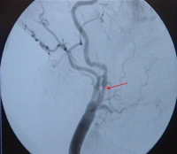
    Digital subtraction angiography (DSA) of the arteries supplying the brain 

    • Only if the noninvasive procedures have proved inconclusive resulting in therapeutic consequences
    • Example: stenotic kinking not evident on MRI or CT scan

    CT or MRI of the brain

    • In symptomatic patients, parenchymal imaging prior to elective revascularization
    • In asymptomatic patients, such imaging can provide important additional insight, e.g., evidence of clinically silent cerebral infarction

    Chest x-ray

    Laboratory panels

    • Blood count
    • Electrolytes
    • Coagulation
    • Kidney function parameters
    • Liver function parameters
    • Blood lipids
    • Blood group

    In all patients with arteriosclerotic carotid stenosis, other sequelae of arteriosclerosis (coronary artery disease [CAD], peripheral arterial occlusive disease [PAOD]) should be assessed!

  4. Special preparation

    • Mark affected side
    • Have patient fast 2–6 hours as per department protocol
    • Continue platelet inhibitors; in combined anticoagulation, decide case-by-case based on cardiac risk profile
    • Determine blood group
  5. Informed consent

    • Puncture site: hematoma; pseudoaneurysm; AV fistula; infection
    • Vascular injury from balloon dilation -> syncope, hemiparesis; if stenting fails -> emergency open surgery
    • Hyperperfusion syndrome -> possibly cerebral hemorrhage
    • Change of puncture site
    • Allergic reaction to contrast media, drugs, latex
    • Stent displacement, possibly, surgical revision
    • Vascular dissection/perforation e.g., catheter induced
    • Allogeneic blood transfusion with risk of infection (hepatitis, HIV)
    • Bleeding risk due to fibrinolysis
    • HIT II following heparin injection
    • contrast-agent induced nephropathy, possibly lifelong dialysis
  6. Anesthesia

    • Preferably local anesthesia
    • General anesthesia if requested by patient or when cooperation is lacking
  7. Positioning

    PM 317-2

    Supine, arm abducted on side of puncture ( the DSA unit is advanced from the contralateral side!), head reclined and turned to contralateral side

  8. Operating room setup

    PM 317-3

    The surgeon stands on the side to be operated on, with the assistant facing him/her. The scrub nurse stands next to the assistant towards the patient’s feet. The C-arm for intraoperative angiography approaches from the contralateral side, with the monitor positioned next to the patient's head and clearly visible to the surgeon.

  9. Special instruments and fixation systems

    • Mobile or stationary DSA unit or hybrid operating room
    • High pressure injection pump for contrast medium
    • Various sheaths(6F and larger)
    • Standard instrument set for vascular surgery, retractor
    • Various guidewires (Terumo® long, at least 180 cm, soft-tipped stiffer wires specifically for carotid dilation).
    • Various guiding catheters (such as RDC = Renal Double Curve, Hockey Stick, SIM 1 and 2 = Simmons type sidewinder)
    • Dilation balloons in different sizes (3–6 mm)
    • Balloon-expandable stents specially designed for the carotid artery (4–6 mm, length 20–30 mm, straight and conical, e.g., for co-treatment of common carotid artery)
    • Instrument set ready for conversion to open vascular surgery, if intervention fails or complications are imminent
    • In case of increased risk of embolization possibly protection device

    Caution:

    1. Protection device: Any exploratory maneuver poses an embolic risk per se. Without protection two probings are needed, with protection at least three. No protection device is employed in the video case because it involved scar-induced restenosis.

    2. Carotid interventions require longer wires and catheters than interventional procedures on the iliac axis or abdominal aorta.

  10. Postoperative management

    Postoperative analgesia:

    Follow this link to PROSPECT (Procedures Specific Postoperative Pain Management) and to the current German guideline Behandlung akuter perioperativer and posttraumatischer Schmerzen [Treatment of acute perioperative and posttraumatic pain].

    Postoperative care

    • Monitoring on Intermediare Care Unit for 24 hours
    • Cardiovascular monitoring, esp. blood pressure
    • Laboratory panels
    • Neurological checks: postop. on day of surgery and also on day 2–3
    • Color flow Doppler imaging from day 3 postop.
    • ASA 100 1x1 , possibly in combination with clopridogel

     Deep venous thrombosis prophylaxis

    • Usually low molecular weight heparin s.c. perioperatively every 24 hours    

     Ambulation

    • After 24 hours

    Physical therapy

    • Not required

    Diet

    • Starting 4 hours post surgery

    Bowel movement

    • Not required

    Work disability

    • Around 2 weeks