Start your free 3-day trial — no credit card required, full access included

Perioperative management - Exstirpation of a solitary parathyroid adenoma

  1. Indications

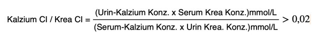
    Any laboratory chemical detection of a primary hyperparathyroidism (pHPT)
    Urgent surgical indication after hypercalcemic crisis

    • The term pHPT describes by definition the primary autonomous hypersecretion of parathyroid hormone from one or more parathyroid glands (PTG). It must be distinguished from the secondary form as a result of an underlying disease associated with hypocalcemia (vitamin D deficiency, chronic renal insufficiency).
       
    • A pHPT occurs sporadically or hereditarily, usually in the context of multiple endocrine neoplasia (MEN).

    Pathophysiology

    The hormonal control of calcium levels is primarily regulated by parathyroid hormone (PTH, synthesis in the parathyroid glands) and vitamin D3 (1,25-dihydroxycholecalciferol, synthesis of the active metabolite in the proximal tubules of the kidneys). The secretion of PTH by the parathyroid glands is inversely regulated. A slight increase in serum calcium concentration and the associated activation of the CaSR result in inhibition of PTH secretion. Conversely, a low calcium concentration stimulates the synthesis and secretion of PTH. In patients with pHPT, the normal regulation of PTH secretion is disrupted. Despite an elevated serum calcium concentration, these patients exhibit disproportionately high PTH levels and suffer primarily from the consequences of the resulting hypercalcemia.

    Epidemiology

    In pHPT, a solitary parathyroid adenoma is found in 85 - 90% of cases, women are affected 3 - 4 times more frequently than men. The disease is usually diagnosed between the ages of 55 and 75.

    This must be distinguished from multi-gland disease, which usually has a genetic cause. These hereditary forms must be identified or distinguished. Hereditary primary hyperparathyroidism (hpHPT) often affects multiple glands either as diffuse hyperplasia or in the form of multiple adenomas.

    About 2 - 5% of all pHPT cases occur in the context of multiple endocrine neoplasia type 1 (MEN 1) with equal frequency in women and men at an early age.

    pHPT is the leading diagnosis in MEN 1 and is observed almost regularly. Although MEN 4 affects the same primary organs as MEN 1 (parathyroid glands,pancreas and pituitary gland), patients usually present later in life and have a more indolent disease course than patients with MEN 1. Most patients with MEN 4 and hyperparathyroidism have single parathyroid adenomas, while in MEN 1 all four parathyroid glands are hyperplastic. MEN 4 is much rarer than MEN 1.

    In MEN 2a with leading tumor medullary thyroid carcinoma, pHPT occurs in 15 - 10%.

    In < 1 % of cases, there is a parathyroid carcinoma: Often younger patients with severe pHPT, association with syndromic diseases, e.g. familial HPT-jaw tumor syndrome: This disease is characterized by severe pHPT and tumors in the jaw bones. In this disease, the PTG tumors are malignant in 15 - 30% of cases.

    The presentation of pHPT has changed in the last three decades from a highly symptomatic disease to an incidentally discovered hypercalcemia, particularly due to the easy availability of laboratory diagnostics. The diagnosis of pHPT is made at least 10 years earlier today than 20 years ago.

    Surgery

    Surgery is the only curative therapy; practically all patients with proven pHPT benefit from surgery. Due to the morbidity spectrum of hypercalcemia, parathyroidectomy is indicated not only in symptomatic forms but also in asymptomatic cases as a rule.

    Preoperative detection of a single enlarged parathyroid gland enables a focused surgical approach.

    The standard procedure for solitary adenoma is the open minimally invasive parathyroidectomy demonstrated here as the initial procedure for localized pHPT, usually combined with histological frozen section examination and intraoperative PTH monitoring. If the adenoma has been localized preoperatively concordantly with ultrasound and scintigraphy, intraoperative monitoring of the PTH level can be omitted.

    Through a 2 - 3 cm long skin incision, the affected parathyroid gland is removed in a focused manner. The procedure takes place exclusively in one thyroid lobe. Due to the anatomical location, resection of an upper parathyroid gland is more difficult than that of a lower one. If the adenoma is not found or the PTH level does not drop, bilateral exploration must be performed and all four parathyroid glands identified.

    In case of suspicion of a carcinoma, possibly extension by en-bloc resection of adjacent structures (hemithyroidectomy, straight neck muscles, soft tissue). Lymphadenectomy only if suspicious lymph nodes are detected.

  2. Contraindications

    • cardiopulmonary risk assessment
       
    • General anesthesia incapacity
       
    • Coagulation disorder or intake of anticoagulants

      • Guidelines recommend before elective operations an individual benefit-risk analysis: If the operative bleeding risk clearly outweighs the potential cardiovascular benefit, the ASA therapy should be discontinued.
      • In cases of higher-grade anticoagulation such as P2Y12-ADP receptor antagonists (e.g. Clopidogrel), NOACs (e.g. Xarelto) or Vitamin K antagonists (e.g. Falithrom or Marcumar), an interdisciplinary council regarding the indication for anticoagulation, possibility of bridging with heparin, and operative bleeding risk should develop a therapy concept
         
    • The rare disease of familial hypocalciuric hypercalcemia (FHH) should be considered in the differential diagnosis for all patients with asymptomatic pHPT, as parathyroidectomy is not helpful in FHH, the hypercalcemia persists. See also under Diagnostics/Differential diagnoses
       
    • Conservative therapy with Cinacalcet, if surgery is not feasible for anesthesiological reasons or in patients who, despite detailed counseling, do not want to undergo surgery. Cinacalcet binds to the calcium-sensing receptor (CaSR) and thus leads to reduced PTH synthesis and secretion and thereby to a lowering of the calcium level.

      • Note: In case of surgical feasibility, Cinacalcet should never be used permanently instead of a first intervention.
         
    • In case of negative localization diagnostics or suspicion of multiglandular disease -> bilateral cervical exploration
  3. Preoperative Diagnostics

    Symptoms

    The classic symptom triad of "stone, bone, and stomach pain" in the sense of nephrolithiasis, osteitis fibrosa cystica, as well as dyspepsia or gastric/duodenal ulcers is rarely found today. Often, the suspicion of pHPT is raised due to incidentally detected elevated serum calcium levels. This development of pHPT into an oligo- to asymptomatic disease is at least partially attributed to the routinely performed automated determination of serum electrolytes, including serum calcium, since the mid-1970s.

    The symptoms of pHPT are primarily determined by hypercalcemia

    • Kidney stones and nephrocalcinosis, which arise from the increased calcium excretion in the urine.
       
    • Bone pain, osteoporosis, pathological fractures.

      • PTH causes increased bone resorption and thus leads to osteoporosis in the long term. Bone pain is a typical symptom. Bone lesions with greatly reduced density are referred to as so-called "brown tumors" or osteofibrosis cystica. These advanced findings are rarely observed nowadays.
         
    • Peptic ulcers in the stomach and duodenum

      • Gastrin-induced gastric juice secretion in the context of hypercalcemia or MEN syndromes (gastrinomas, Zollinger-Ellison syndrome)
         
    • acute pancreatitis
       
    • In early diagnosis, neurocognitive and psychiatric symptoms such as fatigue, lack of drive, weakness in concentration up to depression and anxiety states are in the foreground. These symptoms are initially not perceived as an illness, but often attributed to advanced age and should definitely be further investigated for pHPT if hypercalcemia is detected.
       
    • Calcification of the vessels with cardiovascular consequences

    Laboratory Diagnostics

    • In principle, the diagnosis of pHPT is made based on pathological laboratory parameters: hyperparathyroidism with consecutive hypercalcemia with normal renal function.

      Note: Only then is localization diagnostics justified.
       
    • To exclude a coincidence of pHPT and thyroid autonomy
      -> Determination of TSH, fT3/T4
       
    • further laboratory diagnostics (phosphate, alkaline phosphatase, albumin/total protein;

    Differential Diagnoses

    • Familial hypocalciuric hypercalcemia (FHH)
      For differential diagnosis, in mild pHPT or postoperatively persistent pHPT, the syndrome of familial hypocalciuric hypercalcemia (FHH) must be considered. FHH is an autosomal dominant inherited disorder caused by a heterozygous inactivating mutation in the calcium-sensing receptor (CaSR) with lifelong hypercalcemia and relative hypocalciuria. Parathyroid hormone is inappropriately high, measured against calcium as an expression of PTH resistance. The phenotype is asymptomatic, there is no indication for surgery.

      Note: With elevated PTH value and high serum calcium level, pHPT is clearly present only if the calcium excretion in the urine is not reduced.
    Diagnosis of Primary Hyperparathyroidism
    Laboratory Chemistry
    Serum calcium (best ionized or albumin-corrected): elevated
    Parathyroid hormone: elevated
    Serum phosphate: reduced
    Renal values: in the normal range (if elevated: elevated parathyroid hormone)
    Vitamin D level: in the normal range (if reduced: elevated parathyroid hormone)
    Alkaline phosphatase: indication of increased bone remodeling


    Formel.png
    Conc concentration, Crea creatinine, Cl clearance


    • Exclusion of FHH by determining the calcium and creatinine concentration in serum and in 24-hour collected urine. After determining the calcium clearance / creatinine clearance quotient (ratio) (CCCR > 0,02), FHH can be excluded.
       
    • Vit D deficiency

      Compensatory increase in PTH due to calcium deficiency.

      Note: With high PTH value and normal calcium level, there is almost always a vitamin D deficiency or renal insufficiency.
       
    • Medication intake

      Long-term therapy with lithium can lead to HPT typically as a multi-gland disease.
       
    • Hypercalcemia in paraneoplastic syndrome

      Due to the release of a protein related to parathyroid hormone (PTHrP) by the tumor.
       
    • Hereditary primary hyperparathyroidism (hpHPT)

      In patients ≤30 years with confirmed pHPT, based on a detailed family and personal history, after genetic counseling, a mutation analysis of potential candidate genes should be performed.

    Localization Diagnostics

    • Ultrasound is the localization diagnostic tool of first choice, with which about 70% of parathyroid adenomas can be diagnosed. While normal-sized parathyroids are not visible, adenomatously altered parathyroids usually appear as homogeneous, solid, and hypoechoic structures. Malignancy criteria exist if the parathyroid is >3cm or poorly demarcated from the thyroid capsule and its surroundings.
      At the same time, the thyroid gland, which is useful in this context, can also be examined. There may be a coincidence of pHPT with thyroid pathology (thyroid autonomy, nodular goiter, etc.), which influences the surgical strategy.
       
    • Results as good as with ultrasound can be achieved with Tc-99m-methoxyisobutyl-isonitrile (MIBI) scintigraphy. The informative value of scintigraphy can be further improved by applying SPECT technology. SPECT (Single Photon Emission Computed Tomography) combines the gamma camera with computed tomography and thus enables three-dimensional imaging of the compartment to be examined. In contrast to ultrasound, scintigraphy can also depict mediastinal adenomas.
       
    • The sensitivity of computed and magnetic resonance tomography is reported in the literature as 30 – 80 % and is thus below that of ultrasound and scintigraphy.

    Note: A negative localization diagnostic correlates with lower surgical success, so further localization methods or primary bilateral cervical exploration should be discussed with the patient. The probability of multi-gland disease increases from 5 - 10 % to about 20 %.

    Imaging Reserve Procedures

    They are available only in a few centers and are therefore rarely used in initial imaging diagnostics. They can be helpful for localizing a parathyroid adenoma before a recurrence procedure, but should always be indicated in weighing against a high hit rate in exploration by an experienced surgeon.

    • 4D CT/MRI

      The relatively new variant of 3D imaging is supplemented by the additional "dimension" of different contrast medium perfusion of tissue over time. Pathological parathyroids can be visualized through differences in their wash-out behavior of the contrast medium compared to the surroundings.
       
    • PET-CT/MR (C-11-methionine- or F-18-fluorocholine-PET-CT/MR)

      The depiction of parathyroid adenomas with various nuclides such as C-11-methionine and F-18-fluorocholine in PET-CT or PET-MR can further improve preoperative localization. The accuracy is around 90 - 95 %, but is offered only in a few centers and is not a service of statutory health insurance. Therefore, these procedures are recommended only before recurrence procedures.
       
    • Selective venous catheter and superselective angiography for regionalization of PTH secretion have only minor importance, possibly as an intraoperative procedure for ectopic parathyroid adenomas.
       
    • Fine-needle aspiration cytology (FNAC) with determination of PTH in the aspirate, especially in suspicion of an intrathyroidal parathyroid adenoma. In case of suspicion of malignancy, the examination is contraindicated.
  4. Special Preparation

    Tools for histological and biochemical success control intraoperatively should be available:

    • Frozen section examination: Can confirm parathyroid tissue, but cannot distinguish between hyperplasia and adenoma.
       
    • Intraoperative peripheral-venous parathyroid hormone determination (IOPTH). The IOPTH is considered the best quality indicator for postoperative normocalcemia. 10-15 min after adenoma removal, the drop in PTH is measured. For intraoperative PTH measurement, an established interpretation criterion and a standardized protocol should be used. Common are both the "soft" Miami criteria with a drop in PTH level after adenoma removal to < 50 % of the baseline value, as well as the "hard" Halle criteria with a drop in PTH level < 35 pg/ml. With the Miami criteria, there is a risk of overlooking a multiglandular disease, with the Halle criteria the risk is greater of unnecessarily exploring the contralateral side.

      Note: If the PTH drop does not meet the required criteria, bilateral exploration must be performed, also considering ectopic localizations. If the adenoma was preoperatively localized consistently by 2 imaging procedures, the intraoperative PTH determination can be dispensed with.
       
    • Pre- and postoperative laryngoscopy
       
    • Pre- and post-resection vagus stimulation using neuromonitoring. In intubation anesthesia, intraoperative neuromonitoring is preferably performed using a tube electrode to derive the M. vocalis electromyogram. Verifiable documentation of the stimulation EMG of the ipsilateral vagus before and after resection is recommended for forensic reasons.
      LINK to PDF IONM
       
    • Antibiotic prophylaxis only for operation duration > 3h and depending on the risk profile (diabetes, immunosuppression, obesity, dialysis requirement, severe underlying disease)
  5. Informed Consent

    • usual risks of a surgical procedure (bleeding, infection, wound healing disorder, follow-up procedure)
       
    • failure to locate and revision procedure
       
    • persistent hyperparathyroidism
       
    • postoperative hypoparathyroidism
       
    • recurrent laryngeal nerve palsy
       
    • autotransplantation into neck or forearm muscle
       
    • exploration of the thyroid gland with possible resection in case of abnormalities
       
    • pneumothorax
       
    • if necessary, postoperative calcium substitution
       
    • recurrence
  6. Anesthesia

    Intubation anesthesia
    Use of short-acting muscle relaxants only during anesthesia induction, avoidance during the IONM phase

    In suitable patients, the procedure can be performed under local anesthesia.

  7. Positioning

    Positioning
    • Supine position with reclined head (so-called struma positioning)
       
    • both arms adducted
  8. OR Setup

    OR Setup

    The surgeon stands contralateral to the side being operated on. The first assistant stands opposite the surgeon. The scrub nurse stands next to the assistant towards the foot end.

  9. Special Instruments and Holding Systems

    • Thyroid retractor
       
    • Neuromonitoring equipment, also vessel loop for retracting the vagus nerve
       
    • Titanium clips
       
    • recommended: loupes for the surgeon
  10. Postoperative Treatment

    postoperative Analgesia: Parathyroid interventions are among the procedures with low pain intensity, therefore a standard therapy with non-steroidal anti-inflammatory drugs is recommended. Follow the link here to PROSPECT (Procedures Specific Postoperative Pain Management)
    Follow the link here to the current guideline: Treatment of acute perioperative and posttraumatic pain

    medical follow-up treatment: 

    • Close clinical monitoring in the first 4 to 6 postoperative hours to detect/exclude postoperative bleeding.
    • On the first postoperative day, routine check of serum calcium and PTH.
    • Prophylactic temporary substitution of calcium and calcitriol to avoid clinically relevant hypocalcemias, beware of Hungry Bone Syndrome (see Complications), recommended. Standard substitution over 1-2 weeks with 1-3 g calcium daily orally plus 1-2x 0.25ug calcitriol, followed by tapering dosage under clinical and laboratory monitoring.
    • After initial operation of a hpHPT, check calcium and PTH every 3-6 months in the first year, then once per year.
    • Removal of the Redon drainage, if inserted, on the first postoperative day.
    • Laryngoscopy before discharge.

    Thrombosis prophylaxis: In the absence of contraindications, due to the medium thromboembolism risk (surgical procedure > 30min duration), in addition to physical measures, low-molecular-weight heparin should be administered in prophylactic, possibly weight- or disposition risk-adapted dosage until full mobilization is achieved.
    To be noted: Renal function, HIT II (history, platelet count)
    Follow the link here to the current guideline: Prophylaxis of venous thromboembolism (VTE)

    Mobilization: immediate

    Physiotherapy: none

    Diet buildup: immediate

    Stool regulation: if necessary, laxatives from the 2nd day

    Inability to work: 1 - 2 weeks