Start your free 3-day trial — no credit card required, full access included

Restthyroidectomy with partial central lymph node dissection on the left

  1. Skin Incision/Scar Excision

    Video
    Skin Incision/Scar Excision
    Soundsettings

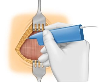
    The operation demonstrates a completion surgery following a right hemithyroidectomy performed years ago due to papillary thyroid carcinoma. The current findings revealed a left uninodular goiter with an approximately 7 mm centrally located nodule and perithyroidal lymph node enlargement on the left.

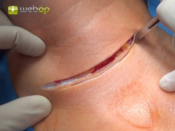
    Excision of the old scar from a previous Kocher's collar incision.

  2. Mobilization of Wound Edges

    Mobilization of Wound Edges
    Soundsettings

    Mobilization of a skin-platysma flap cranially up to the larynx and caudally to the jugulum, with care to preserve the straight cervical veins as much as possible.

  3. Entering the Thyroid Bed at the Anterior Edge of the Sternocleidomastoid Muscle

    Entering the Thyroid Bed at the Anterior Edge of the Sternocleidomastoid Muscle
    Soundsettings

    Fig. right (1) Sternocleidomastoid muscle, (2) Sternothyroid muscle, (3) Sternohyoid muscle

    Since a right hemithyroidectomy had already been performed, the straight neck muscles are not split longitudinally in the midline as usual, but atypically, the entry into the thyroid bed is made immediately at the anterior edge of the left sternocleidomastoid muscle. For better visualization of the thyroid bed in this procedure, the omohyoid muscle is transected. During the course of the preparation, the surgeon switches sides to the left.

  4. Exposure, Mobilization, and Neuromonitoring of the Vagus Nerve

    Exposure, Mobilization, and Neuromonitoring of the Vagus Nerve
    Soundsettings

    Identification of the vagus nerve between the internal jugular vein and the common carotid artery. Mobilization of the nerve and neuromonitoring is performed.

  5. Identification of the Recurrent Laryngeal Nerve

    Identification of the Recurrent Laryngeal Nerve
    Soundsettings

    The recurrent laryngeal nerve is visualized on the dorsal side of the left thyroid lobe after careful dissection, confirmed by the use of neuromonitoring.

    Note: If the nerve cannot be visualized, the use of neuromonitoring is highly recommended. Initially, the vagus nerve should be stimulated, confirming the regular conduction function of the nerve loop (vagus nerve via the recurrent laryngeal nerve to the vocal muscle). Subsequently, systematic palpation of the retrothyroid space with the stimulation probe until a corresponding acoustic signal is audible and a typical electromyographic curve is visible on the monitor.

    Tips for Exposure, Dissection, and Preservation of the Recurrent Laryngeal Nerve

    Tip 1: Atraumatic Nerve Exposure
    At the suspected location, connective tissue fibers are carefully lifted with atraumatic forceps, and the nerve is exposed with a delicate dissecting clamp, such as a fine Overholt clamp, whose branches are to be guided parallel to it, with spreading movements.

    Tip 2: Surgical loupes
    The use of surgical loupes is helpful during dissection. The approximately 1 to 2 mm thin nerve is recognizable by its whitish structure with typical vascular markings.

    Tip 3: Variability of Nerve Course
    If the nerve is not found at its typical location in the esophagotracheal angle, it may be displaced anteriorly from its regular anatomical position by the dislocation of the thyroid, especially if it runs through a vascular bifurcation of the inferior thyroid artery. Special caution is advised in cases of recurrent goiter, and the use of neuromonitoring is therefore highly recommended.

    Tip 4: Anatomical Variant

    With a frequency of approximately 1%, a non-recurrent laryngeal nerve is found. Here, too, the nerve is not found in its typical location but runs horizontally, or also ascending or descending from the vagus nerve directly to the larynx.


  6. Partial Resection of the Ipsilateral Central Lymph Node Compartment

    Partial Resection of the Ipsilateral Central Lymph Node Compartment
    Soundsettings

    For better mobilization of the left thyroid lobe, the straight neck muscles are detached from the anterior surface of the thyroid and held medially with a small hook.

    Then, preparation is done from caudolateral of the left thyroid lobe towards the medial, where one encounters the described significantly enlarged lymph nodes centrally. Here, a lymph node conglomerate with surrounding adipose tissue is carefully dissected out after identifying the dorsally located recurrent laryngeal nerve and sent for frozen section examination.

    Note: Later, there is no indication of malignancy.

Visualization of the Recurrent Laryngeal Nerve to the Larynx

During the dissection of the retrothyroid space, the lower left parathyroid gland cannot be reliabl

Activate now and continue learning straight away.

Single Access

Activation of this course for 3 days.

US$9.30  inclusive VAT

Most popular offer

webop - Savings Flex

Combine our learning modules flexibly and save up to 50%.

from US$7.19 / module

US$86.33/ yearly payment

price overview

general and visceral surgery

Unlock all courses in this module.

US$14.38 / month

US$172.70 / yearly payment