Start your free 3-day trial — no credit card required, full access included

Restthyroidectomy with partial central lymph node dissection on the left

Reading time readingtime 51:26 min.
  1. Skin Incision/Scar Excision

    Video
    Skin Incision/Scar Excision
    Soundsettings

    The operation demonstrates a completion surgery following a right hemithyroidectomy performed years ago due to papillary thyroid carcinoma. The current findings revealed a left uninodular goiter with an approximately 7 mm centrally located nodule and perithyroidal lymph node enlargement on the left.

    Excision of the old scar from a previous Kocher's collar incision.

  2. Mobilization of Wound Edges

    Video
    Mobilization of Wound Edges
    Soundsettings

    Mobilization of a skin-platysma flap cranially up to the larynx and caudally to the jugulum, with care to preserve the straight cervical veins as much as possible.

  3. Entering the Thyroid Bed at the Anterior Edge of the Sternocleidomastoid Muscle

    Video
    Entering the Thyroid Bed at the Anterior Edge of the Sternocleidomastoid Muscle
    Soundsettings

    Fig. right (1) Sternocleidomastoid muscle, (2) Sternothyroid muscle, (3) Sternohyoid muscle

    Since a right hemithyroidectomy had already been performed, the straight neck muscles are not split longitudinally in the midline as usual, but atypically, the entry into the thyroid bed is made immediately at the anterior edge of the left sternocleidomastoid muscle. For better visualization of the thyroid bed in this procedure, the omohyoid muscle is transected. During the course of the preparation, the surgeon switches sides to the left.

  4. Exposure, Mobilization, and Neuromonitoring of the Vagus Nerve

    Video
    Exposure, Mobilization, and Neuromonitoring of the Vagus Nerve
    Soundsettings

    Identification of the vagus nerve between the internal jugular vein and the common carotid artery. Mobilization of the nerve and neuromonitoring is performed.

  5. Identification of the Recurrent Laryngeal Nerve

    Video
    Identification of the Recurrent Laryngeal Nerve
    Soundsettings

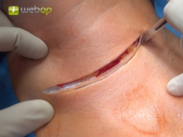
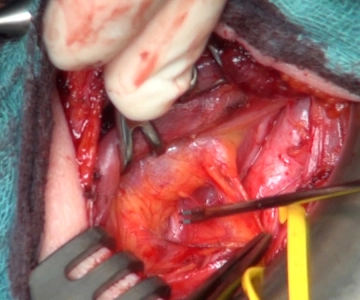
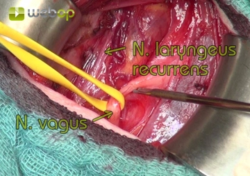
    The recurrent laryngeal nerve is visualized on the dorsal side of the left thyroid lobe after careful dissection, confirmed by the use of neuromonitoring.

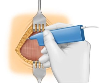
    Note: If the nerve cannot be visualized, the use of neuromonitoring is highly recommended. Initially, the vagus nerve should be stimulated, confirming the regular conduction function of the nerve loop (vagus nerve via the recurrent laryngeal nerve to the vocal muscle). Subsequently, systematic palpation of the retrothyroid space with the stimulation probe until a corresponding acoustic signal is audible and a typical electromyographic curve is visible on the monitor.

    Tips for Exposure, Dissection, and Preservation of the Recurrent Laryngeal Nerve

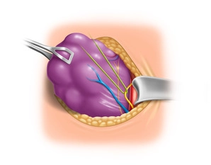
    Tip 1: Atraumatic Nerve Exposure
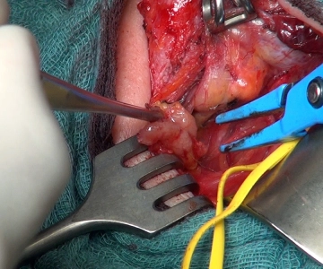
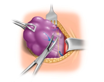
    At the suspected location, connective tissue fibers are carefully lifted with atraumatic forceps, and the nerve is exposed with a delicate dissecting clamp, such as a fine Overholt clamp, whose branches are to be guided parallel to it, with spreading movements.

    Tip 2: Surgical loupes
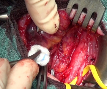
    The use of surgical loupes is helpful during dissection. The approximately 1 to 2 mm thin nerve is recognizable by its whitish structure with typical vascular markings.

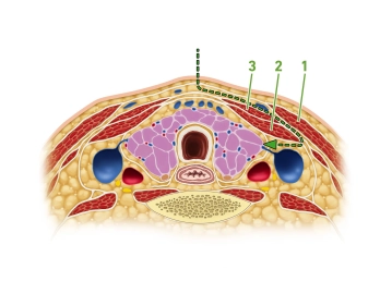
    Tip 3: Variability of Nerve Course
    If the nerve is not found at its typical location in the esophagotracheal angle, it may be displaced anteriorly from its regular anatomical position by the dislocation of the thyroid, especially if it runs through a vascular bifurcation of the inferior thyroid artery. Special caution is advised in cases of recurrent goiter, and the use of neuromonitoring is therefore highly recommended.

    Tip 4: Anatomical Variant

    With a frequency of approximately 1%, a non-recurrent laryngeal nerve is found. Here, too, the nerve is not found in its typical location but runs horizontally, or also ascending or descending from the vagus nerve directly to the larynx.


  6. Partial Resection of the Ipsilateral Central Lymph Node Compartment

    Video
    Partial Resection of the Ipsilateral Central Lymph Node Compartment
    Soundsettings

    For better mobilization of the left thyroid lobe, the straight neck muscles are detached from the anterior surface of the thyroid and held medially with a small hook.

    Then, preparation is done from caudolateral of the left thyroid lobe towards the medial, where one encounters the described significantly enlarged lymph nodes centrally. Here, a lymph node conglomerate with surrounding adipose tissue is carefully dissected out after identifying the dorsally located recurrent laryngeal nerve and sent for frozen section examination.

    Note: Later, there is no indication of malignancy.

  7. Visualization of the Recurrent Laryngeal Nerve to the Larynx

    Video
    Visualization of the Recurrent Laryngeal Nerve to the Larynx
    Soundsettings

    During the dissection of the retrothyroid space, the lower left parathyroid gland cannot be reliably identified, and macroscopically, no structure corresponding to a parathyroid gland adheres to the thyroid capsule. The nerve is now successively traced cranially along the dorsal thyroid capsule to its insertion into the larynx. No scar tissue changes are found here, indicating that only the right side was actually operated on at that time.

  8. Interruption of the Upper Pole Vessels

    Video
    Interruption of the Upper Pole Vessels
    Soundsettings

    Mobilization of the upper thyroid pole by selectively sealing and transecting the branches of the superior thyroid artery, partially using clips.

    Caution: With a high-reaching pole, there is a risk of injury to the external branch of the superior laryngeal nerve if the vessels are not transected close to the thyroid. Consideration should be given to using EBSLN monitoring (External Branch of the Superior Laryngeal Nerve)! 

  9. Visualization of the Upper Parathyroid Gland

    Video
    Visualization of the Upper Parathyroid Gland
    Soundsettings

    The upper left parathyroid gland is identified and carefully dissected from the dorsal thyroid capsule.

    Tips for Preserving Parathyroid Tissue

    Tip1: The following information is important for the localization of the parathyroid glands: The upper ones are located cranial to the inferior thyroid artery and dorsal to the recurrent nerve. The lower ones are located caudal to the inferior thyroid artery and ventral to the nerve. 

    Tip2: Not only is the identification of the parathyroid gland important, but also its vascular supply. Dissecting the parathyroid gland from the thyroid capsule should be done while preserving a small vascular pedicle if possible.

    Intraoperative fluorescence angiography with indocyanine green or, less elaborately, the use of an autofluorescence identification system can facilitate the detection of parathyroid glands. An assessment of the vascularization or devascularization of the parathyroid gland is only achieved with ICG fluorescence angiography.

    Tip3: If a parathyroid gland has been inadvertently devascularized and is no longer surrounded by vascular-rich connective tissue, autotransplantation is recommended.

    If the upper and lower parathyroid glands are not found intraoperatively, the resected thyroid specimen should be thoroughly inspected on the operating table. Unintentionally removed parathyroid glands are autotransplanted. If a lower parathyroid gland is removed during a central compartment lymphadenectomy for radicality reasons, autotransplantation must be considered.

  10. Mobilization and Extirpation of the Thyroid Lobe

    Video
    Mobilization and Extirpation of the Thyroid Lobe
    Soundsettings

    Using a bipolar clamp, the left thyroid lobe is gradually detached from the anterior tracheal wall and the still partially adherent straight neck muscles, while preserving the recurrent laryngeal nerve. Finally, the thyroid lobe is only attached to the trachea in the area of the Ligamentum Berry (also known as the suspensory ligament of the thyroid gland). After ligating the fine vessels crossing the recurrent nerve with Prolene® 5-0, the thyroid lobe is excised and removed.

  11. Final Neuromonitoring of the Recurrent Laryngeal Nerve and Vagus Nerve

    Video
    Final Neuromonitoring of the Recurrent Laryngeal Nerve and Vagus Nerve
    Soundsettings

    After checking for hemostasis under forced PEEP ventilation, the final neuromonitoring of the recurrent laryngeal nerve and vagus nerve is performed.

  12. Layered Wound Closure

    Video
    Layered Wound Closure
    Soundsettings

    In a dry surgical field, drainage is omitted. The muscle compartment is closed with adapting interrupted sutures, and after releasing the head reclination, a continuous intradermal suture is performed.