Start your free 3-day trial — no credit card required, full access included

Central liver resection (mesohepatectomy) for Klatskin tumor

  1. Complete Liver Mobilization with Exposure of the Sub- and Retrohepatic Inferior Vena Cava

    Video
    Complete Liver Mobilization with Exposure of the Sub- and Retrohepatic Inferior Vena Cava
    Soundsettings

    Upper abdominal right-angle laparotomy and exploration of the abdomen to exclude an extrahepatic tumor and peritoneal carcinomatosis (not shown). Transection of the falciform ligament down to the level of the confluence of the hepatic veins into the inferior vena cava.

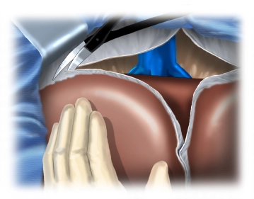
    Release of both liver lobes from adhesions with the triangular ligaments, exposing the retrohepatic inferior vena cava.

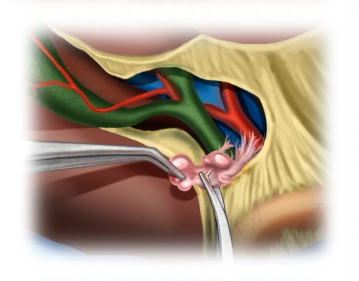
    The inferior vena cava is dissected from caudal to cranial, with the smaller distal hepatic veins being gradually exposed. In this case, two larger, variably occurring inferior venous branches are initially transected between Overholt clamps and ligated.

    The management of the short central hepatic veins into the inferior vena cava is performed depending on the caliber, using either a clip or transfixion ligature.

    165-D-5

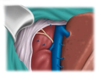
    Tip: Generous retrohepatic mobilization facilitates resection and prevents blood loss or injury to the vena cava during anterior traction of the liver during parenchymal transection.

  2. Visualization of the Ligamentum hepatoduodenale with complete extrahepatic lymphadenectomy

    Visualization of the Ligamentum hepatoduodenale with complete extrahepatic lymphadenectomy
    Soundsettings

    Beginning of the preparation of the Lig. hepatoduodenale, in which the gallbladder is released anterogradely. Then a complete dissection of the hepatic hilum is performed with removal of all connective and lymphatic tissue.

    Presentation and looping of the left and right hepatic artery, both of which are not affected by the tumor, as well as the ductus hepatocholedochus.

    Note: The right hepatic artery undercrosses the main bile duct, and the left hepatic artery originates atypically from the left gastric artery.

  3. Preparation for Left Lateral Liver Transection by Dissection Along the Left Portal Vein Branch

    Soundsettings

    A prerequisite for resection is that the right hepatic artery, which runs immediately behind the bile duct bifurcation, is not affected by the tumor. Likewise, tumor-free status of both portal vein branches must be ensured. Another requirement is that the remaining liver volume exhibits optimal function to ensure adequate postoperative liver function.

    Dissection along the left portal vein branch into the liver fissure. After transecting a parenchymal bridge, the gap between the left lateral lobe (Segment 2/3) and Segment 4 opens at the lower end of the umbilical fissure. The left portal vein trunk running here is exposed, and all branches leading medially to Segments 4a and 4b are transected, as were the Segment 4 arteries previously. The portal vein branch to Segment 1 is isolated and managed.

    Then transection of the bile duct with rapid section and smear collection, removal of the bile duct stent, and suture closure of the distal stump.

    Note: An intraoperative rapid section examination is recommended to aim for an R0 resection. BilIN (biliary intraepithelial neoplasia) may be accepted in case of doubt. However, there is considerable uncertainty here due to discontinuous tumor and perineural sheath growth.

  4. Looping of the Left Hepatic Vein and Hanging Liver Maneuver

    Soundsettings

    Transection of the hepatic veins to segment 1. After dislocating the left liver lobe to the right, isolate and ligate the left hepatic vein with a vessel loop.

    Subsequently, a sling is placed dorsally on the liver between the left and middle hepatic veins to suspend the liver in the resection plane and pull it ventrally (so-called Hanging Liver Maneuver).

    This provides better orientation for the resection plane with the shortest transhepatic path through the parenchyma and simultaneously compresses the transverse intrahepatic blood vessels.

Resection along Segment 2/3 to Segment 4 with Transection of the Middle Hepatic Vein

Marking of the resection boundary between Segment 4 and Segments 2/3 supported by ultrasound (not s

Activate now and continue learning straight away.

Single Access

Activation of this course for 3 days.

US$9.30  inclusive VAT

Most popular offer

webop - Savings Flex

Combine our learning modules flexibly and save up to 50%.

from US$7.19 / module

US$86.33/ yearly payment

price overview

general and visceral surgery

Unlock all courses in this module.

US$14.38 / month

US$172.70 / yearly payment