Start your free 3-day trial — no credit card required, full access included

Perioperative management - Central liver resection (mesohepatectomy) for Klatskin tumor

  1. Indications

    Perihilar/central bile duct carcinoma (Klatskin tumor) originates from the bifurcation of the extrahepatic bile ducts. Due to the almost absent muscular layer of the bile ducts, there is early invasion into the liver parenchyma or the portal vein.

    The only curative treatment option is radical R0 resection, which can only be achieved through an en bloc resection of the bile ducts, bile duct bifurcation with liver resection. The assessment of resectability and development of an appropriate resection strategy is of utmost importance due to the close anatomical relationship to the portal vein bifurcation and the hepatic arteries.

    Curative approaches in advanced stages are regularly associated with significant loss of liver parenchyma and pose a challenge for surgery, anesthesia, and intensive care medicine.

    An isolated resection of the extrahepatic bile duct system (so-called "isolated hilus resection") cannot be considered a curative procedure. The result is local recurrence rates of 70-90% and virtually no long-term survival. The bile ducts of the caudate lobe (Segment 1), which regularly open near the bile duct bifurcation, are predisposed to local recurrences.

    To achieve an adequate safety margin, a simultaneous en bloc resection of the central liver parts together with the bile duct system is always necessary. This includes the caudate lobe as well as the hilar portions of segments 4, 5, and 8. Achieving a proximal and lateral safety margin is often problematic due to the usually diffuse infiltration along the bile ducts and perineural sheaths.

    Extended hemihepatectomies have been established as standard procedures, with right-sided procedures requiring the so-called hilar en bloc resection including the portal vein bifurcation. In left-sided procedures, due to the course of the right hepatic artery, dissection is usually required dorsal to the bile duct, so a general portal vein resection is not indicated here.

    In an inoperable situation due to technical reasons or functional irresectability, liver transplantation is an option, although with poor results so far and a glaring shortage of organs. Improved results are available for selected patients in a multimodal treatment program.

    For these extensive liver resections of an organ damaged by cholestasis and bile duct inflammation, preoperative assessment of residual liver function is essential. Conditioning of the liver through selective bile duct drainage and, before extensive right resections, through hypertrophy induction (portal vein embolization/ALPPS) is an option for improved outcomes.

    Especially when hypertrophy induction is not possible, the procedure described here of central liver resection is a technical variant for radical resection while simultaneously maximizing the preservation of healthy liver parenchyma.

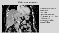
    In the demonstrated case, it is a stenosing bile duct tumor at the hepatic bifurcation Bismuth IIIb, diagnosed by MRCP, ERCP, and brush cytology. An extended right resection was not feasible due to atrophy of the left liver lobe. Therefore, removal of liver segments 1, 4, 5, and 8 was performed while preserving the lateral segments 6, 7, and 2, 3, particularly since the portal vein bifurcation and the left hepatic artery did not appear infiltrated morphologically.

    168-PM1
  2. Contraindications

    Non-resectability of hilar cholangiocarcinoma:

    • Encasement/occlusion of the portal vein
    • Lymph node metastases beyond the hepatoduodenal ligament
    • Distant metastases
    • Malignant ascites
    • Advanced liver diseases, particularly advanced PSC (primary sclerosing cholangitis)
    • Tumors that have reached the segmental bile ducts on both sides simultaneously.
    • Tumors infiltrating both hepatic arteries.

    Liver cirrhosis:

    • Assessing the functional reserve of a cirrhotic liver is challenging. Besides the general physical condition and the Child-Pugh score, the severity of portal hypertension is crucial. The most important parameters for adequate postoperative liver function are normal bilirubin levels and a hepatic venous pressure gradient of < 10 mmHg. Indicators of the extent of portal hypertension include spleen size, the presence of esophageal varices, and platelet count (Caution: < 100,000/μl).
    • Significantly increased risk with portal hypertension due to complicated, risky dissection in the liver hilum.

    Insufficient residual liver volume:

    The most important cause of perioperative mortality after liver resection is liver failure. Risk evaluation is therefore of crucial importance, as therapeutic options for postoperative liver insufficiency are very limited.

    • For Klatskin tumors, a residual liver volume of 40% should be aimed for, as the liver is pre-damaged by cholestasis.
    • Evaluation of hypertrophy conditioning via portal vein embolization (PVE) or ALPPS (Associating Liver Partition and Portal vein ligation for Staged hepatectomy).
    • Corresponding hypertrophy is expected after 3 to 5 weeks, but can take significantly longer in a cholestatic or cirrhotic liver.

    General inoperability:

    Underlying conditions of the patient, particularly cardiac risks, must be considered.

  3. Preoperative Diagnostics

    History: Risk factors include recurrent cholangitis, primary PSC, and bile duct cysts.

    Clinical signs: painless jaundice (itching skin, discolored stool), pain, weight loss, fever

    Tumor markers: Ca19-9, CEA

    Imaging:

    The following diagnostic algorithm is recommended:

    1. (Contrast-enhanced)ultrasound as basic diagnostics

    2. Contrast-enhancedCT of the abdomen

    The standard procedure for surgical planning and clarification of resectability is multiphase contrast-enhanced rapid spiral computed tomography with thin slices. This achieves high-resolution imaging of the arterial, portal venous, and venous structures. With appropriate evaluation software, tumor volume, total liver volume, and remaining liver volume after resection can be estimated. Regarding liver function, the CT provides only indirect indications: enlarged spleen, recanalized umbilical vein, prominent caudate lobe, dilated bile ducts. A disadvantage is inadequate visualization of the bile duct system.

    3. MRI/MRCP

    Before interventional procedures as a non-invasive method for imaging the bile duct system, to assess bile duct conditions without an influencing post-interventional inflammatory reaction. Detached liver segments can also be depicted.

    4. ERCP and/or PTCD possibly with cholangioscopy

    During ERCP, there is the possibility of histological and cytological tissue diagnostics, in specific cases also via EUS-guided fine needle aspiration. ERCP combines diagnostic and therapeutic options in biliary obstruction.

    Tissue sampling is performed in the form of brush cytology and direct forceps biopsy, with the combination of both methods increasing sensitivity.

    A modern cytological method using aspirated bile is FISH (Fluorescence in situ Hybridization) with detection of chromosomal aberrations of the bile duct epithelium.

    A disadvantage is bacterial contamination up to the intrahepatic bile ducts, increasing the rate of infectious complications and potential tumor cell dissemination (liver metastases, implantations during intraoperative removal of the bile duct stent).

    PTCD is usually indicated when ERCP cannot be performed. The advantages of PTCD are more accurate and easily reproducible cholangiography with minimal contamination risk.

    5. Optional staging laparoscopy to exclude liver metastases and/or peritoneal carcinomatosis.

    PET-CT (Positron Emission Tomography combined with CT) 

    Not the first choice in initial diagnostics, as it is inferior to other imaging methods.

    A reasonable indication seems to be the detection of local recurrences after resection of hilar cholangiocarcinomas.

    Endosonography

    Endoscopic ultrasound offers high-resolution imaging of structures adjacent to the stomach and duodenum. This includes the common hepatic duct up to the liver hilum, local lymph nodes, and adjacent vessels. Tissue acquisition is possible via fine needle aspiration (FNA), with sensitivity lower than ERC-based tissue acquisition methods.

    Remark:

    Histological or cytological confirmation of malignancy is only possible in a few cases due to difficult visualization. If there is sufficient clinical suspicion, surgery is indicated even without prior histological confirmation.

    The extent of the tumor disease is ultimately only visible intraoperatively; only then can resectability be determined. Thus, a high exploration rate must be accepted.

  4. Special Preparation

    • Endoscopic drainage before resection:
      • Stents should generally be avoided. However, liver dysfunction increases under cholestasis, which is responsible for increased morbidity and mortality. Therefore, stents are often unavoidable.
      • Before stent placement, the necessary imaging to clarify resectability should be completed. An existing stent also complicates the intraoperative assessment of resectability.
      • Indications: Presence of cholangitis or significant cholestasis, bilirubin level >10 mg/dl, possibly bilateral drainage.
      • In contrast to distal bile duct carcinomas, preoperative decompression of the bile ducts is recommended for hilar tumors. The placement of a bile duct drainage must ultimately be decided by the surgeon, as on one hand, the liver parenchyma remaining postoperatively must be decompressed preoperatively, while on the other hand, cholangiosepsis is one of the main causes of postoperative mortality. After any manipulation of the bile ducts, the risk of bacterial contamination of the bile ducts increases. Therefore, in the case of post-ERCP/stent, antibiotic prophylaxis or therapy before and after liver resection is advised, otherwise single-shot antibiotic is sufficient.
    • Blood group determination with sufficient transfusion reserve (4-6 units of packed red blood cells, possibly FFP or platelets)!
    • In case of increased cardiopulmonary risk, assessment of surgical risk through further diagnostics (stress ECG, cardiac echo, coronary angiography, lung function test)
    • Adequate intensive care capacity for high-risk patients
    • A mild laxative or enema is recommended the day before.
  5. Informed consent

    Standardized consent forms should be used for patient education. These also include appropriate anatomical drawings where findings can be recorded. Alternatives and additional treatment options should be discussed, particularly any extensions of the primary procedure should always be considered.

    General Risks

    • Bleeding
    • Postoperative bleeding
    • Hematoma
    • Need for transfusions with associated transfusion risks
    • Thromboembolism
    • Wound infection
    • Abscess
    • Injury to adjacent organs/structures (stomach, esophagus, spleen, diaphragm)
    • Wound dehiscence
    • Incisional hernia
    • Subsequent surgery
    • Mortality

    Specific Risks

    • Liver parenchyma necrosis
    • Bile fistula
    • Bilioma
    • Hemobilia
    • Biliary peritonitis
    • Bile duct stricture
    • Pleural effusion
    • Air embolism (due to accidental or unnoticed opening of hepatic veins)
    • Portal vein thrombosis
    • Hepatic artery thrombosis
    • Chronic liver failure
    • Liver failure with hepatic coma
    • Tumor recurrence
Anesthesia

Intubation anesthesiaIntra- and postoperative analgesia with epidural catheterFollow this link to P

Activate now and continue learning straight away.

Single Access

Activation of this course for 3 days.

US$9.40  inclusive VAT

Most popular offer

webop - Savings Flex

Combine our learning modules flexibly and save up to 50%.

from US$7.29 / module

US$87.56/ yearly payment

price overview

general and visceral surgery

Unlock all courses in this module.

US$14.59 / month

US$175.10 / yearly payment

to top