Start your free 3-day trial — no credit card required, full access included

Anatomy - Central liver resection (mesohepatectomy) for Klatskin tumor

  1. The Klatskin Tumor

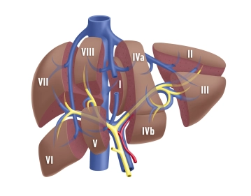
    Klatskin tumors are a subgroup of bile duct carcinomas. The term Klatskin tumor encompasses the entity of perihilar bile duct tumors between the junction of the cystic duct and the branches up to the second generation of the right and left bile duct.

    Etiology
    The current concept for the development of bile duct carcinomas is based, similar to gastrointestinal tract carcinomas, on the so-called adenoma-carcinoma sequence. This is thought to be triggered by a combination of chronic inflammatory changes with increased cell turnover and prolonged or recurrent cholestasis. Predisposing conditions are therefore congenital abnormalities of the bile ducts such as Caroli syndrome, choledochal cysts, abnormal junction of the bile ducts, and inflammatory stenosing bile duct diseases such as sclerosing cholangitis or liver fluke infection.

    Bismuth-Corlette Classification
    Hilar cholangiocarcinomas were first described by Klatskin in 1965. Ten years later, Bismuth et al. classified the ECC (extrahepatic cholangiocarcinomas) according to their anatomical pattern of involvement. This clinical-surgical classification is based solely on the spread of the carcinoma along the bile ducts and distinguishes four main types:

    I. The carcinoma affects only the main bile duct distal to the bifurcation.

    II. The tumor is located in the area of the bifurcation without involvement of the right and left bile ducts.

    III. Both the bifurcation and the right or left bile duct are affected, with IIIa indicating involvement of the right and IIIb indicating involvement of the left bile duct.

    IV. Extensive tumor growth along the bile ducts with involvement of the bifurcation and both bile ducts, including second-order branches or discontinuous tumor growth.

    Regional Lymph Nodes
    The regional lymph nodes of perihilar bile duct carcinoma are only the hilar and pericholedochal lymph nodes in the hepatoduodenal ligament.

  2. Functional Liver Anatomy

    Functional Liver Anatomy

    The liver is macroscopically divided into a larger right and a smaller left lobe (volume ratio approximately 80:20) by the falciform ligament and the insertion of the round ligament of the liver on the diaphragmatic surface, as well as the sagittal fissure on the visceral surface. However, this morphological division does not correspond to the functional structure of the liver. The functional segmentation of the liver is determined by the branching of the portal structures: portal vein, hepatic artery, and bile duct. These three anatomical structures branch not only at the porta hepatis but also predominantly in the same direction within the parenchyma. Each liver segment is completely independent in terms of blood supply and bile drainage from the other segments and can be surgically removed without endangering the function of the remaining liver.

    The term "functional anatomy" thus refers to a substructuring of the liver based on the delineation of hemodynamically independent parenchymal areas, and knowledge of this is essential for the surgical strategy in liver resection procedures.

  3. Portal Vein and Hepatic Veins

    The functional division of the liver is based on the portal branching into individual, independent subunits, the segments.

    Typically, the portal vein divides at the hepatic hilum into a right and left main trunk. The boundary of these supply areas lies along the Cava-Gallbladder line ("Cantlie line"). By further bifurcation of each portal vein trunk, an anteromedial and a posterolateral trunk for liver segments V/VIII and VI/VII are formed on the right side. The left main trunk runs transversely to the left and then as the umbilical part anteriorly, ending at the insertion point of the Ligamentum teres hepatis in the so-called Recessus rex. The left portal main trunk gives off branches for the two left-lateral segments II and III as well as for the median segments IVa and IVb. The caudate lobe holds a special position as it can receive strong inflows from both the left and right portal main trunks.

    According to Couinaud, eight portal venous liver segments are distinguished, numbered clockwise starting with the caudate lobe as Segment I:

    Segment I

    Caudate lobe

    Segment I/II/III

    lateral left liver lobe

    Segment IV

    left paramedian sector (quadrate lobe)

    Segment I/II/III/IV

    left liver half

    Segment V/VIII

    right paramedian sector

    Segment VI/VII

    right lateral sector

    Segment V/VI/VII/VIII

    right liver half

    The liver is traversed in a caudocranial direction by three main venous trunks, namely the right, middle, and left hepatic veins, which divide the liver into a total of four hepatic sectors. The left hepatic vein drains almost exclusively the left-lateral liver lobe and usually joins the middle hepatic vein shortly before its entry into the vena cava, which runs along the Cava-Gallbladder line. The right hepatic vein runs between the posterolateral and anteromedial segments. The caudate lobe has an independent venous outflow, consisting of multiple small veins that drain directly into the vena cava dorsally, known as the Spieghel veins.

    The portal hila of liver segments II, III, and IV are extrahepatic and can be relatively easily dissected in the anterior section of the left umbilical fissure. The hila of the right-sided liver segments are intrahepatic. Exceptions occasionally occur, mostly affecting Segment VI. The anatomy of the hepatic veins is even more variable than that of the portal vein.

    Variants

    Portal Vein System

    • Anomalies of the portal vein bifurcation almost always affect the right portal main trunk
    • Portal vein trifurcation: right main trunk is absent, instead, there are two branches for the right double segments V/VIII and VI/VII (Fig. 1a); occasionally, one of the right branches may also originate from the left portal main trunk (Fig. 1b)
    • Variants of the left portal vein system rarely affect the main trunk, but almost always the division: several small portal vein branches instead of two segmental branches IVa/IVb, occasionally also an additional intermediate branch between the segmental branches II and III
    168_LAV_01_a_b_Pfortader_Lebervenen

    Fig. 1a and 1b: PV = portal vein, LPV = left portal vein, RA = right anterior portal branch, RP = right posterior portal branch

    Hepatic Veins

    • Variants of the hepatic veins are more common than those of the portal vein system
    • Deviations from the hepatic sectors described by Couinaud particularly affect the territories of the right and middle hepatic veins
  4. Hepatic Arteries

    The common hepatic artery originates from the celiac trunk, in rare cases, it originates directly from the aorta or the superior mesenteric artery. After giving off the gastroduodenal artery, the proper hepatic artery divides into the right and left hepatic arteries at the liver hilum. It is not uncommon to find additional extrahepatic branches, such as the artery for segment IV, which usually arises from the left hepatic artery shortly before the umbilical fissure.

    Variants
    • Deviations from the normal distribution type are found in 30% of cases
    • The most common anatomical variants are a proper or accessory hepatic artery from the superior mesenteric artery (Fig. 2a-b) or from the left gastric artery (Fig. 2c-d)
    • Occasionally, both variants are present together (Fig. 2e)
    168_LAV_02_a_b_Leberarterie
    168_LAV_02_c_d_Leberarterie
    168_LAV_02_e_Leberarterie
  5. Bile Ducts

    The extrahepatic portion of the Ductus hepaticus sinister is approximately 3-5 cm long and originates in the umbilical fissure from the union of the two ducts from segments II and III. Proximal to this junction, the bile duct from segment IV is usually incorporated.

    The Ductus hepaticus dexter is very short, just under 1 cm, and receives bile secretion via an anterior and posterior branch from segments V, VI, VII, and VIII. Occasionally, the right hepatic duct may be absent.

    Variants

    Ductus hepaticus sinister

    • The most common (approximately 25%) and clinically significant variant is a common opening of the segment IV bile duct with the segment II/III bile ducts (Fig. 3a)
    • In left-lateral resections, corresponding liver donations, and liver splitting, there may be impairments of bile drainage from segment IV
    168_LAV_03_a_Gallenwege

    Ductus hepaticus dexter

    • Occasionally very short or absent, so that the anterior and posterior pedicle for the double segment V/VIII or VI/VII arises directly from the common hepatic duct (Fig. 3b); a typical hepatic bifurcation is then absent
    168_LAV_03_b_Gallenwege

    Fig. 3b to 3g: rp = right posterior bile duct, ra = right anterior bile duct, dhs = Ductus hepaticus sinister

    Opening Variants

    • Opening of the posterior bile duct directly into the cystic duct, common hepatic duct, or choledochus (Fig. 3c-d)
    168_LAV_03_c_Gallenwege
    168_LAV_03_d_Gallenwege
    • Opening of the anterior and much more frequently posterior right bile duct into the left hepatic duct (Fig. 3e-g)
    • Important in left hemihepatectomy: accidental transection can lead to significant bile leaks and segmental cholestasis in the right liver lobe
    168_LAV_03_e_Gallenwege
    168_LAV_03_f_Gallenwege
    168_LAV_03_g_Gallenwege
  6. Regional Lymph Nodes

    The liver has two lymphatic drainage pathways:

    1. For the most part (90%), the lymph from the liver flows to the lymph nodes at the porta hepatis and from there through the Nodi lymphatici coeliaci into the Truncus intestinalis.
    2. The second drainage pathway (10%) involves the superficial area of the Facies diaphragmatica and the Area nuda. The lymph passes through the diaphragm into the Nodi lymphatici phrenici superiores and through mediastinal lymphatic vessels into the right venous angle.