Start your free 3-day trial — no credit card required, full access included

Perioperative management - Complicated incisional hernia repair for a giant parastomal hernia with open, retromuscular bioprosthetic mesh augmentation

  1. Indications

    Indications

    The reconstruction of the abdominal wall in complex incisional hernias or the presence of a laparostoma presents a particular challenge. A "loss of domain" represents the extreme form of a volume shift of the intestines.

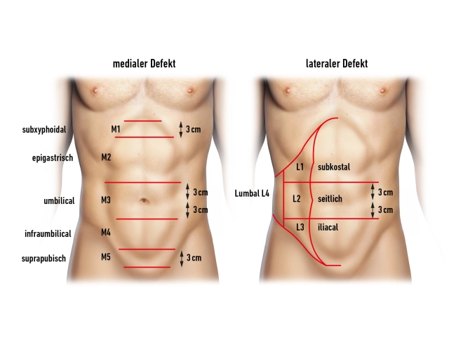
    Complex hernias are considered abdominal wall hernias with defect widths of more than 10 cm, recurrent incisional hernias after mesh implantation (mesh edge herniation, mesh tear, mesh rupture), hernias after flap transfer (denervation, donor site defects), and multiple recurrences.

    In principle, the indication for repair of even a complex hernia is given. Patients lack ventral stabilizing elements of the trunk musculature. Patients with such unstable abdominal walls complain of postural problems with back pain. Physically demanding activities and sports are severely restricted to impossible. An intact ventral abdominal wall is of great importance for physiological processes such as breathing and defecation.

    Furthermore, the abdominal wall defect and the extra-abdominal organ volume will continue to increase. The reconstruction of the abdominal wall ultimately occurs to prevent increasing morbidity.

    The problem with these complex cases is the retraction of the lateral abdominal musculature. The tension towards the midline can be significantly reduced by a length gain of the lateral abdominal musculature.

    There are various techniques for this measure to regain muscle length on the lateral abdominal wall

    1. Abdominal wall relaxation with BTA (preoperative iatrogenic, temporary paralysis of the lateral abdominal musculature with botulinum toxin)
    2. Progressive pneumoperitoneum (abdominal wall dilation through a preoperative pneumoperitoneum)
    3. Intraoperative fascial retraction (traction of the abdominal wall under pharmacological relaxation) see also special preparation

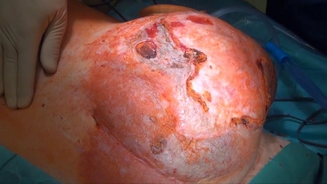
    In the demonstrated case after median laparotomy with sigmoid resection and creation of an end colostomy (Hartmann's situation), a parastomal hernia occurred. Multiple external therapy attempts, most recently with the implantation of a synthetic mesh in onlay technique, led to a progressive incisional hernia recurrence with skin ulceration and partial exposure of the mesh.

    After conditioning the abdominal wall in this case through pneumatic pre-dilation (see abdominal wall reconstruction after Ramirez / Perioperative Management) a renewed hernia repair was performed using the technique described here. The AP relocation offered to the patient with reconnection of the colon was declined.

    Additionally, an abdominal wall component separation was required to achieve a fascial closure in the midline.

    With the anterior component separation ("Ramirez") presented here, midline defects with a width of up to 20 cm periumbilically, up to 8 cm epigastrically, and up to 6 cm suprapubically can be closed by separating parts of the lateral abdominal wall. The technique allows for a tension-free closure with a dynamically competent abdominal wall.

    Due to the extensive detachment of the subcutaneous tissue with the risk of destroying perforating vessels (blood supply to subcutaneous tissue and skin through the deep epigastric vessels) and the resulting high rate of wound healing disorders, hematomas, and seromas, this technique is now considered a second-choice procedure. The posterior component separation, which leads to a release in the area of the transversus muscle, is favored; additionally, the mesh bed can be expanded laterally, dorsally, and retrocostally.

    The reconstruction according to Ramirez was described in the original work without mesh augmentation. The results in the literature favor the simultaneous implantation of a retromuscular mesh.

    With a permanent end stoma, a closure of the parastomal fascial gap according to Sugarbaker with intraperitoneal mesh placement under lateralization of the intestine was additionally performed.

    In infected skin conditions with superficial skin infections and pressure ulcers, biological mesh implants are advantageous and were also used in this situation.

  2. Contraindications

    The indication for hernia repair in patients with liver cirrhosis and ascites should be critically evaluated, and if necessary, preoperative optimization of liver function should be considered. In cases of severe coagulation disorders (Quick < 50%, PTT > 60 s, platelets < 50 /nl) and pronounced portal hypertension with caput medusae, surgery should be avoided, particularly due to the risk of uncontrollable bleeding from abdominal wall vessels.

    It is also important to ensure a good respiratory condition that is not compromised by acute infections. In the presence of respiratory infections, elective surgery should definitely be postponed.

  3. Preoperative Diagnostics

    • The abdominal wall hernia is a clinical diagnosis and can be well recognized in a standing patient. It is advisable to also examine the patient in a relaxed, lying position. When the patient is asked to lift the upper body, the fascial edge, the extent of the fascial defect, and the surrounding muscles can usually be assessed in reducible incisional hernias.
    • For determining the defect location, the extent of the defect, and for depicting the abdominal wall anatomy, CT is the best diagnostic procedure, alternatively an MRI.
    • In cases of previous incisional hernia repairs, a corresponding operative report is often helpful, especially if a mesh repair has already been performed. Here, in addition to the exact surgical technique (extra- or intraperitoneal mesh placement, augmentation or bridging of the fascial defect), the type of mesh material is particularly important.
    • For extensive findings, thorough cardiopulmonary function diagnostics are recommended due to the increase in pressure after repositioning the protruded intestines.

    For better characterization of the present hernia, the EHS classification should be used.

    Classification of primary ventral hernias


    Small (S)

    Medium (M)

    Large (L)


    < 2 cm 

    ≥ 2 - < 4 cm

    ≥ 4 cm 

    Midline

    Epigastric


    Umbilical


    Lateral

    Spigelian


    Lumbar


     Classification of secondary ventral hernias (incisional hernias)

    70-PM-3

    The classification of secondary abdominal wall hernias is initially based on a medial or lateral defect location in the abdominal wall.

    The defect location of medial hernias is then more precisely delineated as subxiphoidal, epigastric, umbilical, infraumbilical, and suprapubic. Laterally, the defects are classified as subcostal, lateral, iliac, and lumbar.

    Further consideration is given to the defect width of incisional hernias: W1 (<4cm), W2 (4-10cm), and W3 (>10cm).

    If multiple hernia defects exist (mesh hernia, Swiss-cheese hernia), their size is determined by the total length and width.

  4. Special Preparation

    • Control of infection situations
    • Medication management in immunosuppression or anticoagulation
    • Control of cardiac and pulmonary risk factors
    • Bowel evacuation is advisable, preoperative bowel lavage is not strictly necessary.
    • Single-shot antibiotic i.v. perioperatively (due to the use of foreign material/mesh) possibly continuation of therapy in case of intraoperative signs of inflammation or bacterial contamination.

    The most important preparation is the assessment of the possibility of repositioning prolapsed organs. In advanced eventration of the intestines, additional conditioning of the abdominal wall is recommended. The following options exist:

    • progressive pneumoperitoneum: Stretching of the abdominal wall by insufflation of air via a catheter into the abdomen. Central venous catheters, small pleural catheters, or similar are used, as no standardized instruments exist. This catheter is pulled through the abdominal wall in a primary procedure. The catheters should be equipped with a bacterial filter and a closure system. Depending on the scheme, room air is applied daily into the abdomen using a large-bore syringe. The preoperative hospitalization of the patient is a disadvantage.
    • Injection of botulinum toxin into the lateral abdominal muscles 4 weeks preoperatively.
    • Intraoperative fascial traction, where the lateral abdominal muscles are mechanically stretched intraoperatively, traction duration 30 min with 14-20 kg.
    • An average length gain of 4-5 cm per side is reported for all methods.

    In the example, conditioning is performed using pneumoperitoneum

  5. Information

    General:

    • Pneumonia
    • Bleeding, rebleeding, hematoma
    • Wound infection/wound healing disorder
    • Thrombosis/embolism
    • Excessive scar formation
    • Injuries to adjacent structures such as nerves, vessels, bladder, and bowel

    Specific:

    • Implantation of synthetic material
    • Reduced load-bearing capacity, especially during the first 3 months
    • Altered shape of the abdominal wall, depressions due to lack of subcutaneous tissue
    • Abdominal wall paralysis
    • Nerve injury/chronic pain
    • Seroma (regularly present, usually without therapeutic consequence)
    • Infection of the implant with the consequence of needing to remove it again.
    • Bowel passage disorder (atony/ileus)
    • Recurrent hernia
    • Bowel perforation
    • Subsequent interventions
    • Lethality
  6. Anesthesia

    • Anesthesia is generally performed under general anesthesia ITN.
    • Intra- and postoperative analgesia using PDK.
  7. Positioning

    Positioning

    Position the patient in a supine position, possibly with one arm positioned alongside.

    Depending on the size of the finding:

    • Slight hyperextension of the patient during preparation or
    • Neutral position during fascial closure
  8. OP Setup

    OP Setup
    • The surgeon stands to the right of the patient.
    • 1st assistant opposite the surgeon, 2nd assistant to the left of the surgeon
    • The scrub nurse stands to the left of the patient next to the assistant.
  9. Special Instruments and Retention Systems

    In the treatment of large, potentially infected incisional hernias, synthetic meshes often prove problematic (mesh ruptures, risk of infection, adhesions). The use of autologous reinforcement techniques (cutaneous flap plasty) has also not been convincing due to the only temporarily ensured stability of the abdominal wall.

    An alternative is biological implants, which are of bovine, porcine, equine, or even human origin. The biological mesh serves as a guide for fibroblast ingrowth and is replaced by the body's own stable and site-specific connective tissue.

  10. Postoperative Treatment

    postoperative analgesia: Non-steroidal anti-inflammatory drugs are generally sufficient; if necessary, an increase with opioid-containing analgesics can be made.
    Follow the link here to PROSPECT (Procedures Specific Postoperative Pain Management).
    Follow the link here to the current guideline: Treatment of acute perioperative and post-traumatic pain.

    medical follow-up treatment:

    • Postoperative monitoring in an intensive care unit or intermediate care unit
    • Single-shot antibiotic therapy in contaminated or infected conditions, prolong if necessary according to antibiogram.
    • After umbilical, incisional, or upper abdominal hernias with mesh implantation, consistent wearing of an abdominal binder for 4-6 weeks is recommended. This is intended to prevent seroma and promote better and faster mesh integration. There is no clear data on this.
    • Redon drains are removed when the output is < 10-20 ml/day.
    • Maintenance of basic mobility and light physical activity. Avoid sports and lifting loads for up to 3 months. Inpatient rehabilitation depending on age, mobility, and home care.

    thrombosis prophylaxis: In the absence of contraindications, due to the medium thromboembolic risk (surgical procedure > 30min duration), low molecular weight heparin should be administered in prophylactic, possibly weight- or disposition-risk-adapted dosage until full mobilization is achieved.
    Note: Renal function, HIT II (history, platelet control)
    Follow the link here to the current guideline: Prophylaxis of venous thromboembolism (VTE).

    mobilization: Immediate

    physical therapy: For large hernias or older patients, intensive respiratory therapy

    dietary progression: Immediate

    bowel regulation: If necessary, oral laxatives from the 3rd/4th day to prevent postoperative bowel atony, constipation should also be avoided in the long term.

    incapacity for work: Depending on the extent of the condition, 3-4 weeks. For occupations with heavy abdominal wall strain → avoid carrying/lifting heavy loads, possibly up to 12 weeks.