Start your free 3-day trial — no credit card required, full access included

Anatomy - diagnostic laparoscopy for adhesion zone, lavage cytology, biopsy sampling, second look operation

  1. Sugarbaker Index (Peritoneal Cancer Index)

    Sugarbaker Index (Peritoneal Cancer Index) 1
    Sugarbaker Index (Peritoneal Cancer Index) 2

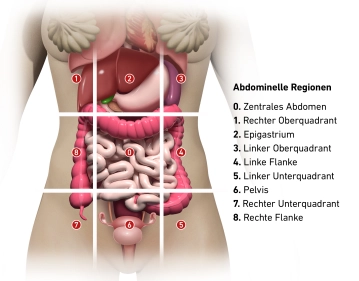
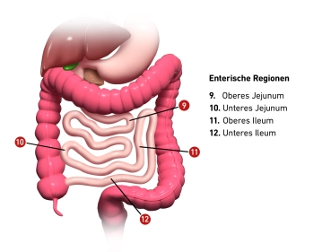
    The Sugarbaker Index, also known as the Peritoneal Cancer Index (PCI), is a scoring system used to determine the tumor burden and distribution in peritoneal carcinomatosis. The PCI divides the abdominal cavity into 13 regions, of which 9 are abdominal and 4 are enteric areas.

    Abdominal regions

    0. Central abdomen

    1. Right upper quadrant

    2. Epigastrium

    3. Left upper quadrant

    4. Left flank

    5. Left lower quadrant

    6. Pelvis

    7. Right lower quadrant

    8. Right flank

    Enteric regions:

    9. Upper jejunum

    10. Lower jejunum

    11. Upper ileum

    12. Lower ileum


    In each of these 13 regions, the size of the largest tumor deposit is assessed and scored as follows:

    - 0 = No visible tumor deposits

    - 1 = Tumor deposits up to 0.5 cm

    - 2 = Tumor deposits 0.5 - 5 cm

    - 3 = Tumor deposits > 5 cm or confluence

    The scores of all 13 regions are then summed to a total score (PCI 0-39), reflecting the extent of peritoneal carcinomatosis.


    RegionOrgans to be assessed
    0 - CentralMidline abdominal incision – Greater omentum – Transverse colon
    1 - Right upperSurfaces of the right liver lobe – Underside of the right diaphragm – Right retrohepatic space
    2 - EpigastriumEpigastric fat pad – Left liver lobe – Lesser omentum – Falciform ligament
    3 - Left upperUnderside of the left diaphragm – Spleen – Tail of the pancreas – Anterior and posterior surfaces of the stomach
    4 - Left flankDescending colon – Left paracolic gutter
    5 - Left lowerPelvic wall lateral to the sigmoid colon – Sigmoid colon
    6 - PelvisFemale internal genitalia with ovaries, fallopian tubes, and uterus – Bladder, Douglas pouch – Rectosigmoid
    7 - Right lowerRight pelvic wall – Appendix
    8 - Right flankRight paracolic gutter – Ascending colon
    9 - Upper jejunum 
    10 - Lower jejunum 
    11 - Upper ileum 
    12 - Lower ileum 
  2. Central

    Greater omentum: The greater omentum is a large, apron-like peritoneal fold that hangs down from the greater curvature of the stomach and covers the transverse colon as well as parts of the small intestine loops. It contains abundant adipose tissue, lymph nodes, and blood vessels and plays an important role in immune defense as well as in isolating infections within the abdominal cavity.

    Transverse colon: The transverse section of the large intestine that runs between the right and left colic flexures. It is located intraperitoneally and is supplied by the transverse mesocolon. The transverse colon is responsible for water absorption and the transmission of intestinal contents to the descending colon.

  3. Top right

    Surfaces of the right liver lobe: The right liver lobe is the largest and functionally most important part of the liver, located in the right upper abdomen. It borders the diaphragm and partially covers the right kidney. The liver plays a central role in metabolism, detoxification, and bile production.

    Underside of the right diaphragm: This structure represents the muscle-tendon plate that separates the abdominal cavity from the thoracic cavity. The right diaphragm is higher than the left and directly borders the right liver lobe. It is involved in respiration by contracting and enlarging the chest cavity.

    Right retrohepatic space: Also known as Morison's pouch, this is a potential space between the right liver surface and the right kidney, clinically significant because fluid can accumulate here in cases of ascites or bleeding.

  4. Epigastrium

    Epigastric fat pad: The adipose tissue in the epigastric region is subcutaneous and protects the underlying organs. It also serves as an energy reserve and insulation.

    Left lobe of the liver: This smaller part of the liver is located in the left upper abdomen and extends across the midline. It is separated from the right lobe by the falciform ligament. The left lobe is anatomically smaller but functionally just as important as the right.

    Lesser omentum: The lesser omentum is a smaller peritoneal fold that connects the liver with the lesser curvature of the stomach and the proximal duodenum. It forms the anterior wall of the omental bursa and contains important structures such as the portal vein, the proper hepatic artery, and the common bile duct.

    Falciform ligament: This structure is a connective tissue attachment that secures the liver to the anterior abdominal wall and the diaphragm. It divides the liver into a right and left lobe and contains the ligamentum teres hepatis, a remnant of the fetal umbilical vein.

  5. Top left

    Underside of the left diaphragm: This region includes the left diaphragm dome, which separates the left pleural field and the left lung from the abdominal organs such as the spleen and the stomach.

    Spleen: The spleen is a highly vascular, intraperitoneal organ located in the left upper abdomen beneath the diaphragm. It has important functions in immune defense, blood filtration, and the breakdown of old or damaged erythrocytes.

    Tail of the pancreas: The tail of the pancreas is located in the left upper abdomen and extends to the splenic hilum. The pancreatic tail is an endocrine and exocrine organ that produces insulin and digestive enzymes.

    Anterior and posterior surfaces of the stomach: The stomach is located in the left upper abdomen and consists of various sections such as the fundus, corpus, and antrum. It is covered by the peritoneum and the omentum from the anterior and posterior abdominal wall. The stomach is essential for the mechanical and chemical digestion of food.

  6. Left Flank

    Descending colon: This section of the large intestine runs along the left side of the abdomen and carries intestinal contents from the transverse colon towards the sigmoid. The descending colon is secondarily retroperitoneal and is involved in water absorption and stool formation.

    Left paracolic gutter: This peritoneal gutter runs lateral to the descending colon and serves as a pathway for the movement of fluids in the abdominal cavity, such as in peritoneal effusions. It forms an anatomical barrier that can restrict pathological processes to one side of the abdomen.

  7. Bottom left

    Lateral pelvic wall of the sigmoid colon: This region includes the lateral pelvic wall, which consists of muscles, fascia, and connective tissue structures that support the sigmoid colon. Nerves and vessels that supply the pelvis and lower extremities also run through here.

    Sigmoid colon: The sigmoid section of the large intestine is S-shaped and connects the descending colon with the rectum. It is located in the lower left abdomen and serves to store and compact feces before they pass into the rectum.

  8. Pelvis

    Pelvis 1
    Pelvis 2

    Ovary (Ovarium)

    The ovary (Ovarium) is located laterally in the pelvic region and runs in a vertical orientation. It has an inner surface facing the pelvic interior (Facies medialis/intestinalis) and an outer surface directed towards the lateral pelvic wall (Facies lateralis). The rounded free edge is called the Margo liber, and at the anterior edge, the Margo mesovaricus, the mesovarium is attached, which is a structure of the broad ligament of the uterus (Lig. latum uteri).

    There are two poles: 

    • Uterine extremity
    • Tubal extremity

    The hilum of the ovary, where vessels and nerves enter, is located at the attachment of the connective tissue of the ovary, known as the mesovarium, at the Margo mesovaricus.

    The ovary is connected to the pelvic wall at the tubal extremity by the suspensory ligament of the ovary. The ovarian vessels (Vasa ovarica), lymphatic vessels, and nerves run along this suspensory ligament.

    The proper ovarian ligament lies between the ovary (uterine extremity). This is located directly behind the tubal angle. The artery R. ovaricus of the uterine artery runs here. 

    The ovary is supplied by: 

    • Ovarian artery
    • Ovarian branch from the uterine artery.

    These arteries form anastomoses at the Margo mesovaricus and supply the ovary with blood. 

    Veins transport the venous blood via the right ovarian vein to the inferior vena cava and via the left ovarian vein to the left renal vein. Part of the blood flows through the uterine plexus to the internal iliac vein.

    The ovary is located in the abdominal cavity (intraperitoneal) in the ovarian fossa. Here, in the retroperitoneum, run the obturator nerve, obturator vessels, and the external iliac vessels. Medially, the umbilical artery and the uterine artery also run.


    The Fallopian Tube (Tuba uterina, Salpinx)

    The Fallopian tube, also known as the Tuba uterina, is about 10-15 centimeters long and 2-5 millimeters thick. It runs intraperitoneally along the upper free edge of the broad ligament of the uterus (Ligamentum latum uteri), from the corner of the uterus to the ovary. In the mesosalpinx, the blood vessels and nerves to the Fallopian tube run, additionally anchoring the tube to the broad ligament.

    The Fallopian tube itself can be divided into different sections:

    • Infundibulum of the Fallopian tube (+ abdominal ostium of the Fallopian tube)
    • Fimbriae of the Fallopian tube, 
    • Ampulla of the Fallopian tube
    • Isthmus of the Fallopian tube
    • Uterine part. 

    The blood supply of the Fallopian tube is provided by small branches (Rr. tubarii) from the ovarian and uterine arteries, which run in the mesosalpinx and anastomose with each other. Veins accompany the arteries and drain into the ovarian vein and the uterine/uterovaginal plexus. 

    The Uterus

    The uterus, also called the womb, is a hollow, muscular organ in the female body that plays an important role during pregnancy and childbirth.

    The uterus consists of:

    • the body (Corpus uteri), 
    • the isthmus uteri
    • the cervix (Cervix uteri) 

    The average length of the uterus is about 7.5 cm, the width 4 cm, and the thickness 2.5 cm. 

    The fundus: uteri represents the upper section of the uterus, located above the entry points of the Fallopian tubes. Its lateral walls are connected to the broad ligament of the uterus (Ligamentum latum uteri). At the lateral edges of the uterus, where the Fallopian tubes enter the uterus, the tubal angle is formed. From the tubal angle, the proper ovarian ligament extends to the ovary, while the round ligament of the uterus extends to the inguinal canal.

    Cervix uteri: The cervix extends with its conical lower third into the vagina. It is divided into the supravaginal portion of the cervix (Endocervix) and the vaginal portion of the cervix (Ectocervix or simply Portio). In the upper area of the vagina, the cervix uteri forms the anterior and posterior vaginal fornices (Fornix vaginae) between the Portio and the vaginal wall.

    Vaginal portion: At the end of the vaginal portion is the external os, also known as the external orifice of the uterus (Ostium uteri or Orificium externum uteri).

    The vaginal portion is usually reddish in color and is covered with stratified, non-keratinized squamous epithelium, similar to the vagina. At the transition to the Ostium uteri, this epithelium changes to the single-layered, columnar epithelium of the cervical canal (Canalis cervicis). Between puberty and menopause, the cylindrical epithelium of the cervical canal can migrate outward onto the Portio (ectropion), leading to more intense redness. With a colposcopy, precancerous stages and cancer can be detected in this area.

    Rectum:

    • Origin of the rectum: Extension of the sigmoid colon, beginning at the upper edge of the third sacral vertebra.
    • Shape and curves:
      • Sacral flexure: Follows the contour of the sacrum downward.
      • Anorectal (perineal) flexure: Bends backward in the area of the perineum, bulging forward.
      • Lateral flexures: Occasionally variable curves in the frontal plane.
    • Structural features:
      • Length of about 15 cm.
      • Three semilunar transverse folds; the most prominent is the Kohlrausch fold about 6 cm above the anus on the right side.
    • Continuation into the anal canal: Ampulla recti above the anal canal, which expands when filled.
    • Boundaries and location:
      • Upper section: Covered by peritoneum in front and laterally, retroperitoneal (Rectum fixum).
      • Peritoneal contact: In women, the peritoneum meets the posterior vaginal fornix and the back of the uterus (Rectouterine pouch) at the middle transverse fold.
      • Below the Kohlrausch fold: Completely extraperitoneal.
    • Anatomical neighborhood:
      • Adjacent posteriorly to the sacrum, coccyx, and the posterior parts of the levator ani muscle.
      • Adjacent anteriorly to the vagina.
    • Vascularization and innervation:
      • Median sacral artery, superior rectal artery.
      • Sympathetic trunks, lateral sacral arteries, sacral plexus in retrorectal connective tissue.

    Urinary Bladder: 

    The urinary bladder is a muscular hollow organ that serves to store urine and later expel it through the urethra. The bladder's capacity is normally 300-500 ml. The bladder can be divided into different parts,

    • the apex of the bladder (Apex vesicae), 
    • the body of the bladder (Corpus vesicae), 
    • the base of the bladder (Fundus vesicae), 
    • the neck of the bladder (Collum or Cervix vesicae), transition to the urethra 

    The apex is connected to the median umbilical ligament (Chorda urachi), the obliterated allantoic duct, which leads to the navel. 

    The peritoneum extends from the bladder to the uterus (Vesicouterine pouch).

    The trigone of the bladder is a roughly triangular, smooth mucosal area located at the base of the bladder. This triangle is formed by the two openings of the ureters (Ostia ureterum) and the internal opening of the urethra (Ostium urethrae internum). The interureteric fold bounds the trigone posteriorly, and anteriorly a corner is formed, which extends into the urethra. In this area, the mucosa is closely fused with the muscularis. In the rest of the bladder, there is a submucosa, which forms a flexible buffer zone between the mucosa and muscle layer, allowing the bladder to fill and empty easily.  

    Douglas Pouch: 

    The peritoneum, also known as the peritoneal membrane, forms a kind of double layer in the pelvic area of the woman, covering and protecting the internal organs.

    The peritoneum lies like a cloth over the uterus. Anteriorly, it covers the bladder and the anterior wall of the uterus, forming a depression at the level of the cervix uteri, called the vesicouterine pouch. It further extends over the fundus and onto the posterior wall to the posterior vaginal fornix. Here, another depression is formed, known as the rectouterine pouch or Douglas pouch. The peritoneum then covers the anterior wall of the rectum. The Douglas pouch is the deepest point in the female pelvis. 

  9. Bottom right

    Right Pelvic Wall: The right pelvic wall consists of a combination of bones, muscles, and fascia that support the pelvis and pelvic organs. Important neurovascular bundles run in this region, supplying the right leg and pelvic organs.

    Appendix: The vermiform appendix is a small, finger-shaped appendage of the cecum. It is located in the right lower abdomen and can become inflamed, leading to appendicitis. The function of the appendix is not fully understood, but it is believed to play a role in the immune system.

  10. Right Flank

    Right paracolic gutter: This structure is a peritoneal groove located lateral to the ascending colon and facilitates the circulation of fluids in the abdominal cavity. It plays a role in the distribution of inflammatory or infectious processes in the abdominal cavity.

    Ascending colon: The ascending part of the large intestine extends from the cecum towards the right colic flexure. It is located retroperitoneally and is involved in the absorption of water and electrolytes to thicken the stool before it is further transported into the transverse colon.

  11. Small intestine

    The small intestine is the longest part of the digestive tract and plays a central role in the breakdown of food and the absorption of nutrients. It measures about 4 to 6 meters and is divided into three sections:

    Duodenum: This section is directly connected to the stomach. Bile from the liver and digestive enzymes from the pancreas enter here, which are necessary to break down fats, proteins, and carbohydrates.

    Jejunum: In the middle section of the small intestine, the majority of nutrient absorption occurs. The inner wall of the jejunum is equipped with tiny protrusions, the villi and microvilli, which increase the surface area and thus maximize nutrient absorption.

    Ileum: The last part of the small intestine primarily absorbs remaining nutrients such as vitamin B12. The ileum finally opens into the large intestine via the ileocecal valve.